


IHC MEGAKARYOCYTES
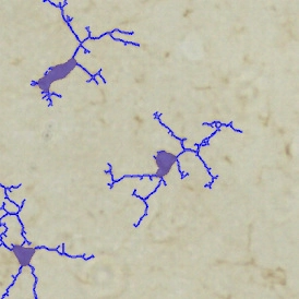
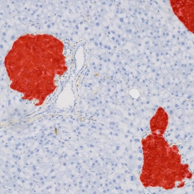
The IHC Megakaryocytes App detects megakaryocytes via marker staining and reports their number, size, and internal neutrophil content.
|
|
|
|


- Overview
- Analysis Steps copy
- Analysis Steps
- Customization
- Output Statistics
- Further Reading
- Downloads
Now Easier Than Ever in StrataQuest 8.0!
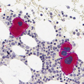
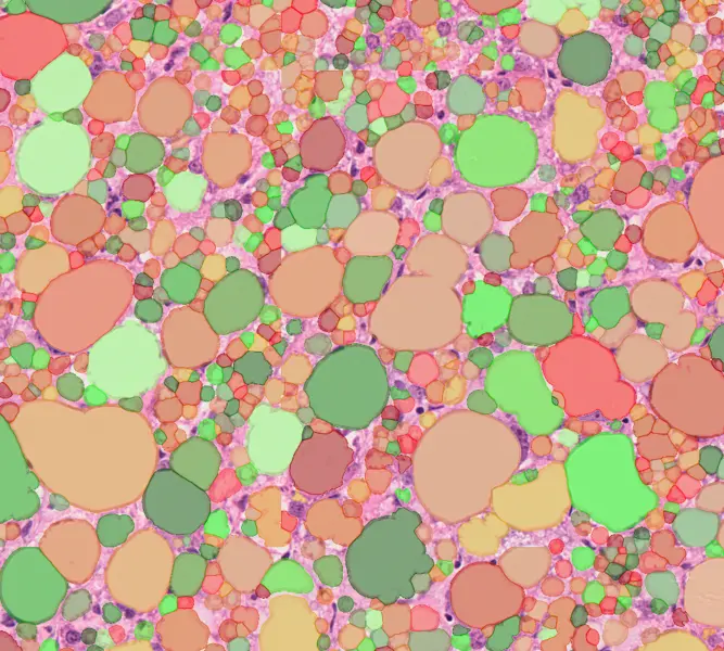
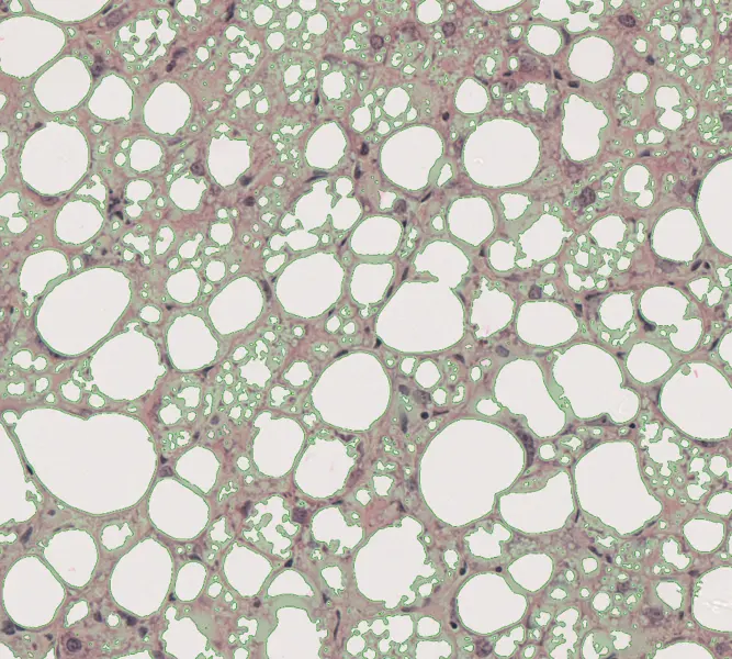
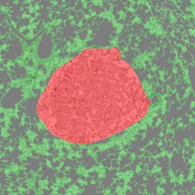
The IHC Megakaryocytes App allows for detection of megakaryocytes based on specific marker staining. It outputs number and size of detected megakaryocytes as well as the number of megakaryocytes that contain neutrophils inside their cytoplasm and the number of neutrophils inside each megakaryocyte. Image courtesy: Prof Wendy Erber and A/Prof Kathy Fuller, The University of Western Australia
|
|
|
|

Customer Publication
13 Sep, 2021
Spatial Neutrophil Distribution in Damaged Skeletal Muscle
News
17 Oct, 2025
Integrative Multiomics Approach Unveils Systemic Dysfunction in Colorectal Cancer (CRC)
Related Apps
IHC LUNG CANCER MOUSE
The IHC Lung Cancer Mouse App uses machine learning to segment tumor and non-tumor regions in murine lung tissue, detect nuclei, and classify cell phenotypes.
Category 3

Custom App development
Perfectly tailored image analysis solutions for your research.
You have a specific research question that needs to be answered? We offer custom development of image analysis pipelines for specific tasks, be it detection of cellular phenotypes or quantification of tissue structures. After discussing your goals with one of our experts, you will get a ready-to-use App and be a step closer to an impactful publication.