Explore the
Tissue Landscape
Whole-slide imaging and analysis solutions tailored to your research
Unlock cellular insights
Visualize and assess phenotypes and spatial distribution across entire slides.

Advanced Imaging Made Simple
Our modular and scalable whole slide imaging systems are built to evolve with your research, offering high image quality and precision.

Support You Can Count On
Collaborate with a committed team of experts for tailored solutions, from pipeline optimization to unique customization.

Powerful Data Exploration
Explore complex datasets with integrated dimensionality reduction tools to uncover relationships and drive meaningful conclusions.

Smart Image Analysis Tools
Use predefined pipelines and advanced image analysis options to tackle even the most complex research questions.
Explore Our Imaging and Analysis Solutions
Latest News

White Paper
13 May, 2026
A New Cas9 mRNA‑Based Therapy Holds Promise to Treat Muscular Dystrophy
Quantitative confocal imaging helps researchers to confirm muscle protein restoration after Cas9 mRNA delivery via targeted lipid nanoparticles.
Blog Post
07 Apr, 2026
How immunofluorescence image analysis factors into NSCLC studies
It’s crucial to understand how immunofluorescence image analysis factors into NSCLC studies to uncover spatial biomarkers and guide research.
White Paper
30 Mar, 2026
Understanding NeuroCOVID-19: SARS-CoV-2 Disrupts Astrocyte Homeostatic Functions
Quantitative cellular analysis of COVID-19-infected astrocyte cells reveals a more central role of astrocytes in NeuroCOVID-19 and the interruptive impact of SARS-CoV-2 on astrocyte homeostasis capabilities, reframing astrocytes as active protagonists.
Read What Our Customers Are Publishing
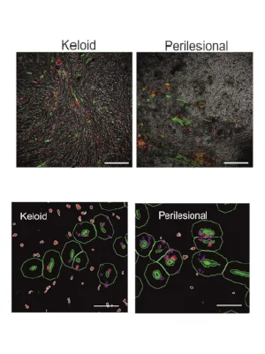
Multi-Antigen Imaging Reveals Key Drivers of Keloid Formation
International Journal of Molecular Sciences
In a recent publication Rath et al (University Hospital Heidelberg) combine multiplexing, image cytometry and in-depth data mining to characterize the immune microenvironment including spatial relationships within keloid lesions. Using this approach, the authors were able to nominate potential therapeutic targets.
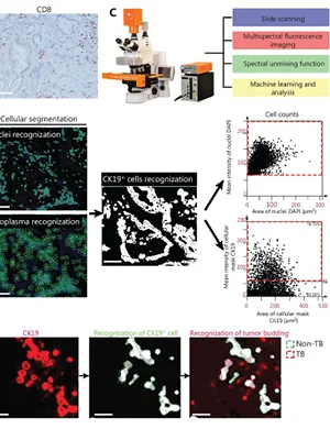
AI-Assisted Analysis of ‘Anti-/Pro-Tumor’ Profiles for Pancreatic Adenocarcinoma
Cancer Biology & Medicine
To predict survival of patients with pancreatic adenocarcinoma, Zhou et al. performed an AI-assisted comprehensive analysis of CD8+ T cells, cancer stem cells (CSCs), and tumor budding (TB). They used StrataQuest image analysis software to automatically detect and quantify tumor-infiltrating CD8+ T cells, CD133+ CSCs, and CK19+ TB.
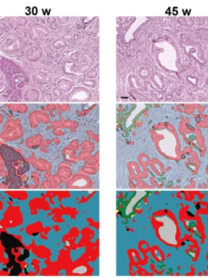
COL8A1 Promotes Tumor in Pancreatic Ductal Adenocarcinoma
Matrix Biology
Yan et al. investigated several collagen subtypes involved in pancreatic ductal adenocarcinoma desmoplasia. Based on this study, COL8A1 was suggested as a new marker for predicting poor chemotherapy responsiveness and survival. The TissueFAXS imaging platform and StrataQuest analysis solution were used to image and segment morphological structures.
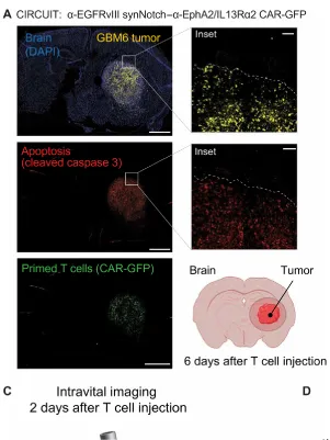
SynNotch-CAR T cells overcome challenges in treating glioblastoma
Science Translational Medicine
A recent publication from the group of Choe et al. (University of California, UCSF) published in Science Translational research is exploring the therapeutic effectiveness of SynNotch-CAR T cells in a PDX glioblastoma mouse model. T cells in-situ were imaged/assessed by TissueFAXS and quantified via StrataQuest.
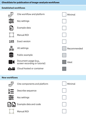
Community-developed checklists for publishing images and image analyses
Nature Methods
QUAREP-LiMi is an initiative focused on promoting quality assessment and reproducibility for instruments and images in light microscopy. Dr. Rupert Ecker, CEO of TissueGnostics, participated in developing standardized guidelines for best practices in image communication. These checklists offer key recommendations on image formatting and annotation among others.
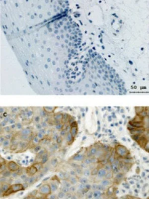
Erk1/2-Dependent HNSCC Cell Susceptibility to Erastin-Induced Ferroptosis
cells
Savic et al. investigated the Erk 1/2 pathway role in ferroptosis induction by targeting xCT protein during the treatment of HNSCC. They found erastin to be a potent ferroptosis inducer which is strongly dependent on Erk 1/2 kinases. TissueFAXS and HistoQuest were used to image the IHC-stained samples and calculate the percentage of xCT-positive cells.
P2X7 receptor isoform B is a key drug resistance mediator for neuroblastoma
Frontiers in Oncology
Arnaud-Sampaio et al. addressed the persistent problem of drug resistance in cancer and elucidated the role of the P2X7 receptor in chemoresistance, using TissueFAXS platform for image acquisition and StrataQuest software for in-depth cytometric quantifications. The paper was published in Frontiers in Oncology.
Cytotoxic CD8+ T cells may be drivers of tissue destruction in Sjögren’s syndrome
scientific reports
In this study, published in scientific reports Nature, Kaneko et al. investigated the role of CD4+ and CD8+ T cell subsets in salivary glands of patients with Sjögren’s syndrome. In order to perform an in-depth quantitative analysis, the power of TissueFAXS platform, and both TissueQuest and StrataQuest software were utilized.
Checkpoint Molecules Predict Response to Immunotherapy in Cancer
cells
Heij et al. investigated the expression of checkpoint molecules in the tumor microenvironment of intrahepatic cholangio-carcinoma. They found that co-expression patterns of checkpoint molecules such as PD-L1 and TIGIT correlate with metastases and poor prognosis. TissueFAXS and StrataQuest were used to image the mIF-stained samples and assess protein expression.
Temporal changes in T cell subsets and expansion of cytotoxic CD4+ T cells in the lungs in severe COVID-19
Clinical Immunology
Kaneko et al. published a study in the Clinical Immunology journal focusing on the quantitative analysis of T-cell subset alterations in thoracic lymph nodes and lung samples of COVID-19 patients. With the help of TissueFAXS and StrataQuest, the authors were first to show the expansion of cytotoxic CD4+ T cells in the lungs of patients with severe COVID-19.
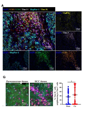
Spatial and functional analysis of Tim3/Tim3-Ligands in tumor development
Molecular Therapy
In a recent paper in Molecular Therapy, Wang et al (Cheeloo Medical College of Shandong University) combine multiplexing and TissueGnostics’ image cytometry solutions for the assessment of the expression level of Tim-3, a well-known immune checkpoint, in tumor infiltrating lymphocytes, which the study identified as a potential target for cancer immunotherapy.
CpG Methylation Influence on Protease Serine 3 Isoforms in Lung Cancer
Acta Pharmaceutica Sinica B
Lin et al. investigated the UHRF1/DNMT1–MZF1 axis in the context of CpG methylation of protease serine 3, a protein with several isoforms that show paradoxical effects. To differentiate the isoforms, the researchers employed 7-marker multiplex staining and performed a quantitative analysis using TissueFAXS SPECTRA and StrataQuest.
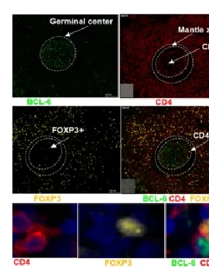
Impact of HIV on T Follicular Regulatory Cells in Human Lymph Nodes
BMC Immunology
Mahlobo et al. published a study in BMC Immunology addressing the effect of HIV infections on frequencies, function, spatial localization and heterogeneity of T follicular regulatory cells (TFRs) in human lymph nodes. TFRs within lymph node tissues were quantified using TissueFAXS i PLUS and TissueQuest.
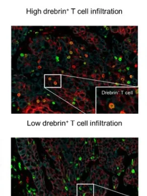
Characterization of the Spatial Immune Context of Lung Cancer
International Journal of Molecular Sciences
In a recent study, Imamura et al. investigated the role of debrin-expressing tumor infiltration lymphocytes (TIL) in lung cancer. The high-end image analysis solution, StrataQuest, was used for the automatic tissue classification and localization of CD3+/debrin+ cellular phenotypes in the tumor cell nest, as well as in the stroma.
Case Report: Tumor Microenvironment Characterization of Rectal Cancer
Frontiers in Immunology
Song et al. investigated the genetics and tumor microenvironment of a rectal cancer patient with MSS/PD-L1-negative recurrent hepatopulmonary metastasis. TissueFAXS and StrataQuest were used to image stained tissue samples and to assess tumor infiltrating immune cells using, among others.
Interested in Learning More?
Contact us and our experts we’ll be in touch soon.