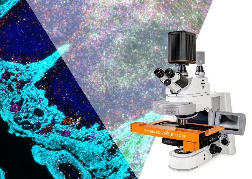
TissueFAXS Platform
Slide scanners designed to adapt to your research today and grow with your discoveries tomorrow.
Software Suite 8 is here — DNN tissue detection, label capture with existing cameras, and a redesigned interface with smarter, more intuitive tools.
See what’s new in Version 8 ›

Modular Imaging Platform
Built for your research needs.
The TissueFAXS platform includes a full family of modular scanning systems - such as TissueFAXS PLUS, i PLUS, Spectra, Q, and SL - engineered to adapt as your research evolves. Configure your system with a wide range of objectives, light sources, filters and cameras, available in upright or inverted configuration. Easily expand to confocal or multispectral imaging, or scale up to 120-slide automation.
- Component Modularity: Objectives (1x–100x), sCMOS cameras, filter sets, light sources, and incubation chamber
- Imaging Modes: Brightfield, fluorescence, confocal, multispectral, and label-free modes (Phase Contrast, DIC, Darkfield, Polarization)
- Automation Scalability: 8 to 120 slides with high-throughput autoloading
- Format flexibility: glass slides, large-format slides, well plates (6–96), chamber slides, and Petri dishes
Why Choose TissueFAXS?
TissueFAXS delivers high-quality images, fast results, and a design that grows with your lab - so you never outgrow your imaging platform.
Modular by Design.
Select only the parts you need now and add advanced features like multispectral or confocal as your experiments grow.
Fits Every Workflow
Choose 8-slide or 120-slide capacity, upright or inverted setup - whatever your lab needs.
One platform. One workflow.
Scan, analyze, and get publication-ready results in one platform.
Ongoing Support You Can Rely On
Our team of experts is ready to help at any stage - before, during, or after setup.
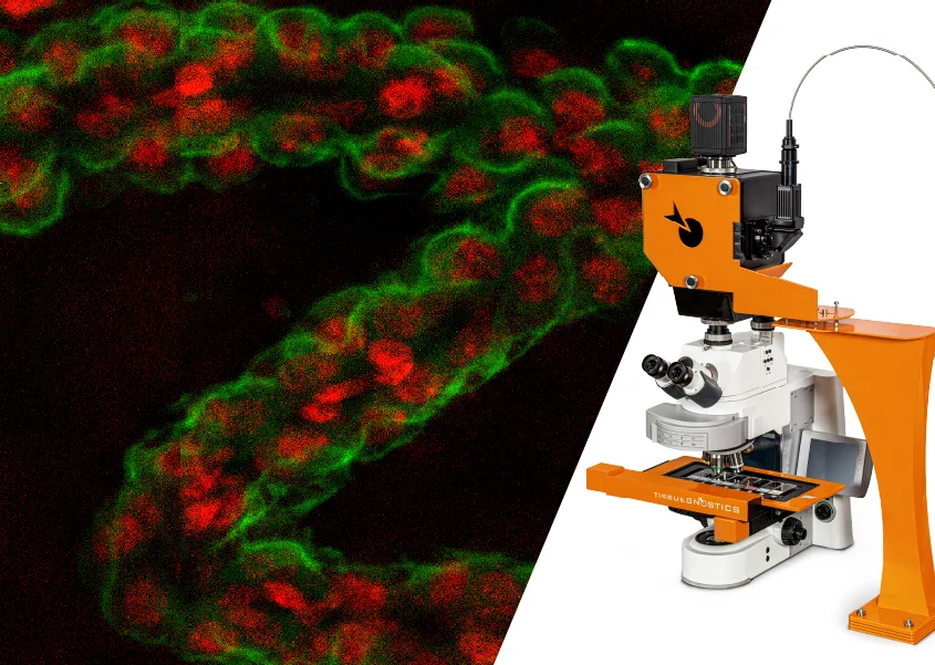
TissueFAXS in Action
High-performance imaging. Scalable systems. Smarter workflows.
Get a quick visual overview of the TissueFAXS platform, its configurations, key features, and how it supports a wide range of imaging workflows.
Which Imaging Configuration Is Right for You?
Understand the strengths and limitations of each setup to find the best fit for your samples and workflows.

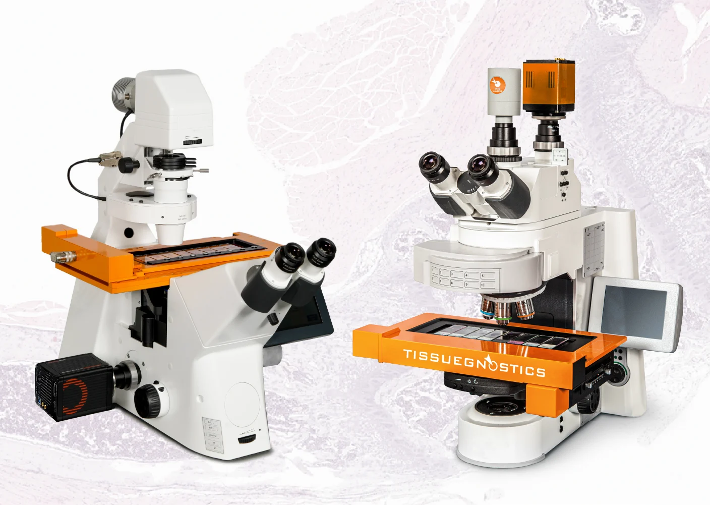
Upright Setup / Top-Down Imaging
- More fluorescence channels for multiplex imaging with an expanded filter wheel
- Fully upgradeable to confocal, multispectral, and high-throughput workflows
- Optimized for slide-based imaging (glass and oversized formats)

Inverted Setup / Bottom-Up Imaging
- More versatility with setup for both slides and plate-based workflows
- Live-cell support with optional incubation module for temperature, CO₂, and O₂ control
- Upgradeable to high-speed confocal (not compatible with multispectral or high-throughput)
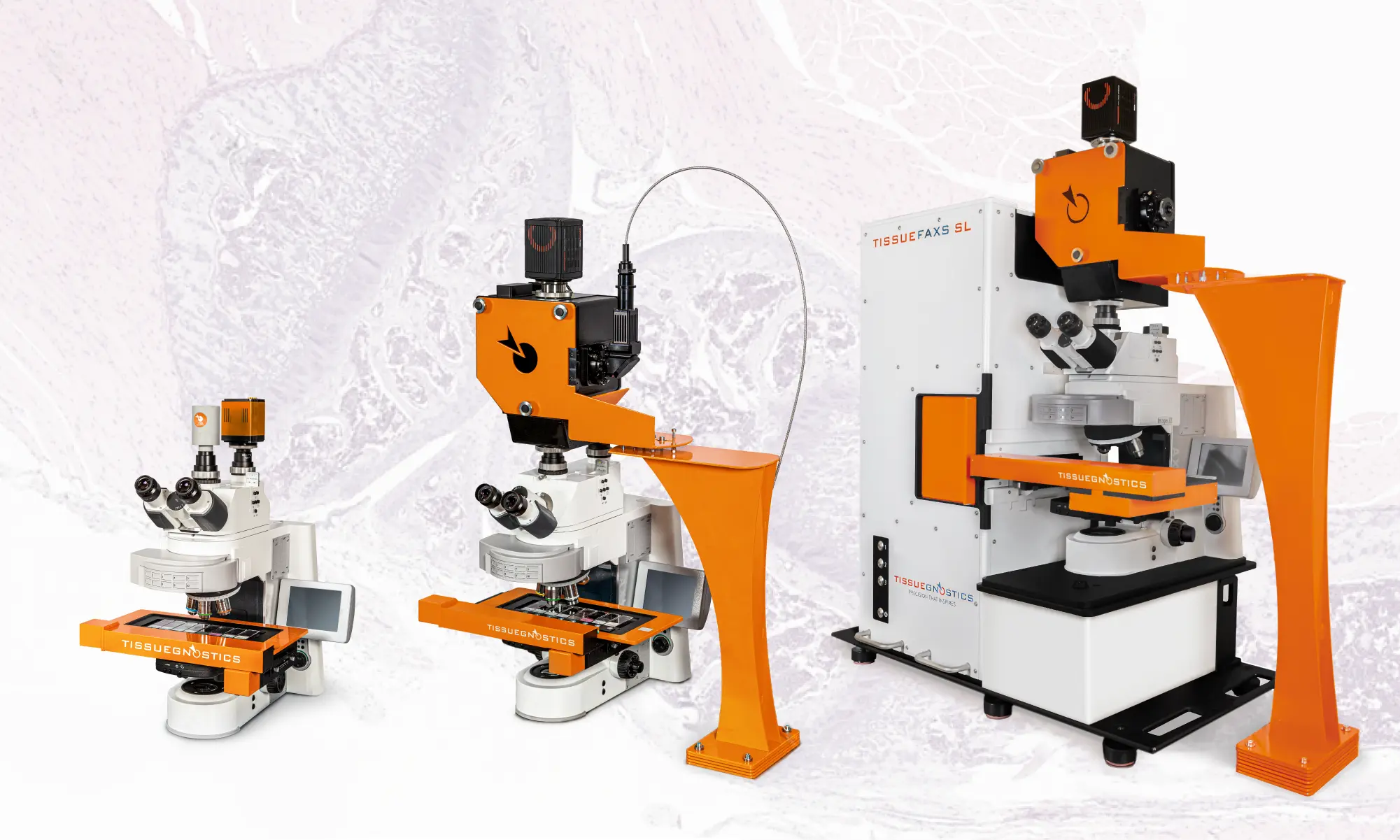
The TissueFAXS Family
Four main configurations for different imaging workflows—all part of one scalable, modular platform.
Multispectral Fluorescence Imaging
TissueFAXS Spectra & Spectra SL
- Detect more markers in a single imaging round with LCTF and lamda stacking technology
- Reduce spectral overlap and bleed-through with spectral unmixing
- Precise unmixing with an extensive reference spectra database
- Ideal for immune profiling, tumor microenvironment studies, and spatial biology
upright
multispectral-imaging
high-throughput imaging
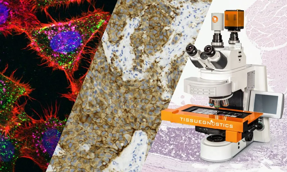
Brightfield & Fluorescence Imaging
TissueFAXS Plus and i Plus*
- Whole-slide imaging in brightfield and fluorescence
- Upright or inverted configurations for tissue and cell culture imaging
- Compatible with oversized slides, well plates, and chamber slides*
- Upgrade-ready for confocal, multispectral, or slide loading
- Designed for research labs needing a flexible, expandable imaging platform
upright
inverted
brightfield
fluorescence
High-Speed Confocal Imaging
TissueFAXS Q, iQ & SLQ
- High-speed spinning disk confocal for fast z-stack acquisition
- Capture crisp, high-resolution images with optical sectioning
- Reduced photobleaching and phototoxicity
- Excellent for thicker samples or subcellular localization
- Used in neuroscience, co-localization, and high-detail tissue studies
upright
inverted
high-speed confocal imaging
high-throughput imaging
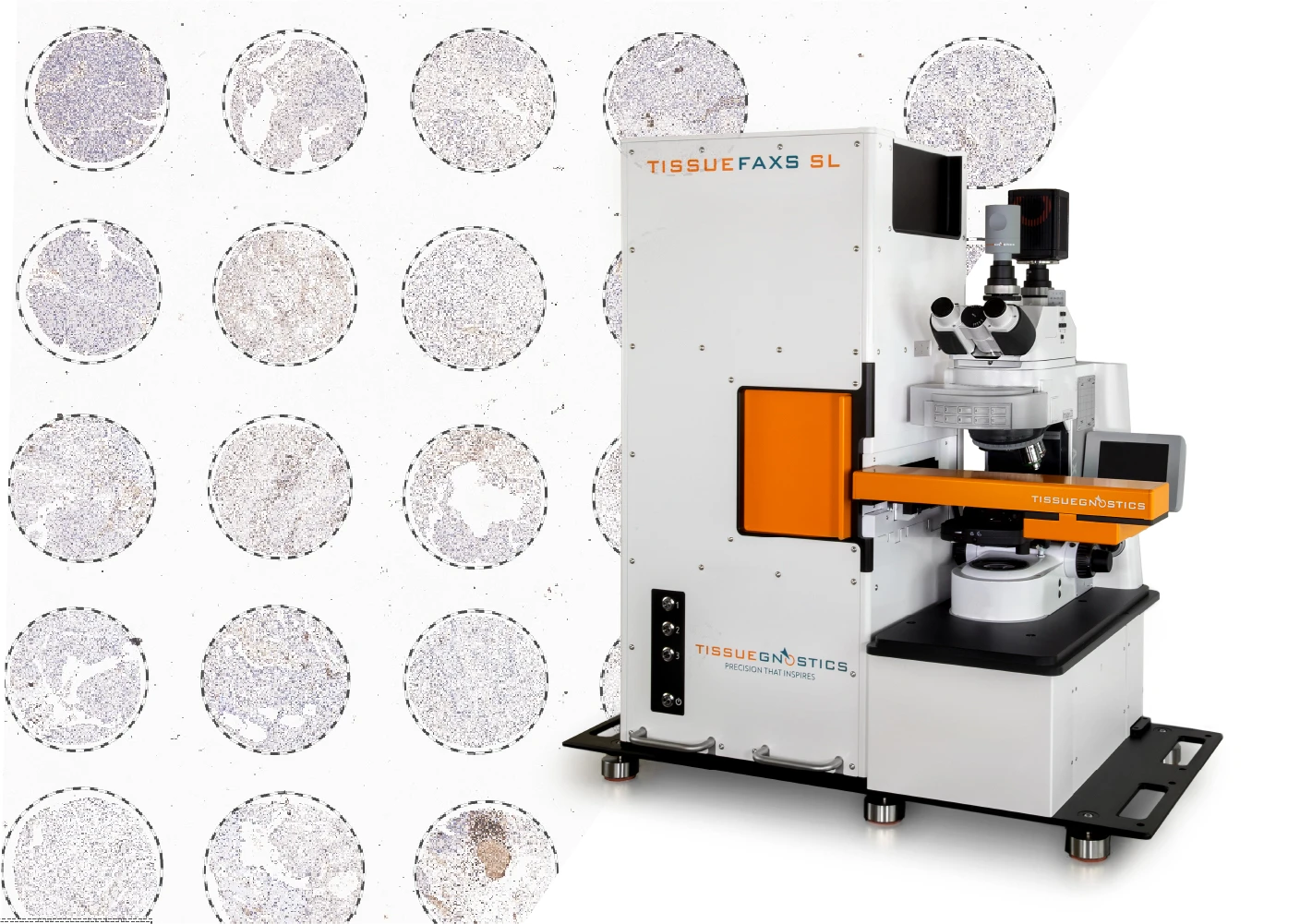
High-Throughput Whole-Slide Imaging
TissueFAXS SL
- Automate batch imaging with up 120-slides per run
- Scan mixed sample types and staining protocols in one batch
- Combine with Multispectral, or Confocal systems
- Ideal for core facilities and large-scale studies
upright
high-speed confocal imaging
multispectral-imaging
high-throughput imaging
Need Help Finding the Right Solution?
Schedule a consultation with our experts and get personalized guidance based on your research goals and workflow needs.
TissueFAXS Upgrade Matrix
Compare base configurations and available upgrade paths.
Configuration | BF | FL | Multispectral Imaging | High-Speed Confocal | High-Throughput | Label-Free Imaging | Inverted Optics | Image Analysis |
|---|---|---|---|---|---|---|---|---|
Base Brightfield & | *Upright Only | Upgrade | *Upright Only | Optional | Optional | Optional | ||
Multispectral Systems | Built-in | *Upright Only | Optional | *Upright Only | Optional | |||
High-Speed | Built-in | *Upright Only | Optional | Optional | Optional | |||
High-Throughput | Upgrade | Upgrade | Built-in | Optional | *Upright Only | Optional |
*Automated slide loaders are only compatible with upright microscopes, as their mechanical design and slide handling require the access provided by an upright setup.
Further Customization Options
Explore modular hardware options to adapt TissueFAXS to your experimental needs, from optics and illumination to filter sets and detection.
- SpectraSplit Filter Set
- Label-Free Imaging
- Objectives 1-100x
- Custom Filter Combinations
- Camera Options
- Light Sources
Optional Filter Set for 7-Marker Imaging
Custom SpectraSplit filters are specifically designed to support simultaneous imaging of up to 7 fluorescent markers with minimal spectral overlap. The system eliminates the need for spectral unmixing by using fixed bandpass filters matched to common fluorochrome excitation/emission spectra (DAPI, Opal Polaris 440, AF488, Cy3, AF594, Cy5, AF750). This approach significantly reduces acquisition time and data size compared to lambda scanning methods while maintaining high signal specificity.


Enhanced Detail Without Stains
Offered as an optional module, TissueFAXS supports Phase Contrast (PC), Differential Interference Contrast (DIC), Darkfield, and Polarization - typically added individually or in pairs based on the user’s application needs. These modes are integrated through components in the optical light path and may occupy one slot in the filter turret, depending on the configuration.
By leveraging natural optical properties like refractive index, birefringence, and light scattering, these techniques reveal fine structural details in live, transparent, or unstained samples. When combined with fluorescence or brightfield scans, they add valuable morphological context for image analysis.
Flexible Magnification for Any Application
TissueFAXS supports most standard ZEISS objectives, allowing configuration with up to seven lenses to suit diverse imaging requirements. The typical setup includes 2.5x, 5x, 20x, 40x (oil), and 63x (oil), with additional options ranging from 1x to 100x in air, water, or oil immersion. This flexibility enables optimal resolution and field of view for applications spanning whole-slide overviews to high-resolution subcellular imaging.

Custom Filter Combinations
Choose from single-, double-, or quad-band filter sets designed to cover a wide range of classical fluorochromes, including DAPI, FITC (Alexa Fluor® 488), Texas Red (Alexa Fluor® 568), Cy3/TRITC, Cy5 (Alexa Fluor® 647), Cy5.5, Cy7, and many more. Our upright microscope configurations support up to 10 filter cubes, while inverted systems accommodate up to 5 filter cubes. This flexibility not only allows precise matching to your experimental needs but also accommodates existing staining protocols, minimizing the need for workflow changes.
Camera Choice for Optimized Imaging
TissueFAXS offers multiple monochrome camera options to match different needs in speed, sensitivity, and resolution. This flexibility supports both high-throughput imaging and low-light fluorescence detection, while also accommodating various budgets.
- PCO edge 4.2LT: 2048 × 2048 px; 40 fps; up to 82% QE
- ORCA Flash 4.0 v3: 2048 × 2048 px; 100 fps; up to 82% QE
- ORCA Fusion BT: 2304 × 2304 px; 89.1 fps; up to 95% QE

Application-Specific Light Sources
TissueFAXS supports a range of solid-state light sources to match different fluorophore sets and imaging demands. Users can select based on spectral range, power output, and independent channel control. Note: Some configurations, especially high-speed or deep multiplexing setups, may require a more powerful source for optimal signal quality.
- Sola SE III: 6-channel white light; 365–735 nm
- Spectra X (nIR): 8 independently controlled LEDs; 380–730 nm
- Spectra III: 8 independently controlled LEDs; 390-750 nm or 390-680 nm; ~500 mW/LED

Browse Additional TissueFAXS Resources
Explore our blog for practical insights, real-world applications, and expert tips on getting the most out of TissueFAXS in your research workflow.

What is a Tissue Slide Scanner?
From multiplex imaging to image analysis, tissue slide scanners help reveal cellular behavior within intact tissues across various research fields.

Investigating CIDs with a Brightfield Microscopy System
Chronic inflammatory diseases (CIDs) involve prolonged inflammation causing tissue damage and dysfunction.

Live Cell Imaging in the Age of AI
When analyzing individual cells, we need technologies that can show us their structures, characteristics, and ongoing processes within the cells.
Related Products

TissueFaxs Imaging Software
For routine workflows and complex imaging tasks.
TissueFAXS features powerful imaging software designed to support both routine and advanced workflows. Users have full access to all scanning parameters allowing protocols to be precisely tailored and saved as reusable profiles for consistent results. Smart autofocus and Z-stacking with a Z-projection (extended focus) algorithm, improve clarity in thick or uneven samples, resulting in high-quality seamless images.
TissueFAXS Viewer
Free. Fast. Made for Collaboration.
TissueFAXS Viewer is a free, standalone desktop application for exploring high-resolution virtual slides generated by TissueFAXS systems. It allows researchers and collaborators to navigate brightfield, fluorescence, or multispectral scans, view annotations, and assess results without needing the full imaging setup. It's the ideal tool for sharing and reviewing data across teams.

Trusted by Thousands of Researchers
Over 3,200 publications using TG solutions
Researchers worldwide rely on TG imaging and analysis platforms to generate high-quality, reproducible data across a wide range of fields—from oncology and immunology to neuroscience and regenerative medicine.

Let’s Build the Right System for You
Tell us about your needs, and we’ll help you find the best configuration - or schedule a personalized demo.
Whether you're imaging slides, well plates, or complex 3D samples, our team is here to help configure a TissueFAXS system that fits your exact workflow. From hardware recommendations to future upgrade planning, we’ll work with you to design a setup that’s ready for now and built for what’s next.