Explore the App Center
Explore our StrataQuest Apps and discover a wide range of biomedical image analysis solutions to inspire your research. If you don’t find the perfect fit for your needs, reach out to us — Our team of application experts are happy to support you by developing custom Apps tailored to your unique analysis requirements.
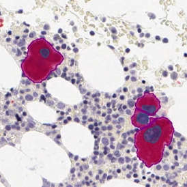
IHC MEGAKARYOCYTES
The IHC Megakaryocytes App detects megakaryocytes via marker staining and reports their number, size, and internal neutrophil content.
bone tissue, bone marrow, megakaryocytes, immunohistochemistry
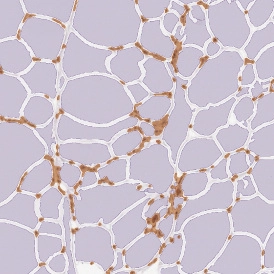
IHC ADIPOCYTES+
The IHC Adipocytes+ App detects adipocytes and cellular aggregates between them. Outputs include number and area measurements for adipocytes and aggregates.
adipocytes, fat tissue, fat cells, immunohistochemistry, immune cell detection
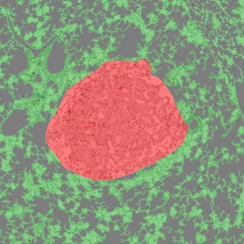
IHC LUNG CANCER MOUSE
The IHC Lung Cancer Mouse App uses machine learning to segment tumor and non-tumor regions in murine lung tissue, detect nuclei, and classify cell phenotypes.
immunohistochemistry, lung cancer, tumor microenvironment, mouse, lung
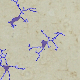
IHC MICROGLIA
The IHC Microglia App detects microglia soma and branching structures, identifying primary and secondary branch points. Outputs include cell count, area, branch length, and branch point numbers.
microglia, central nervous system, peripheral nervous system, phagocytosis, astrocytes, branches
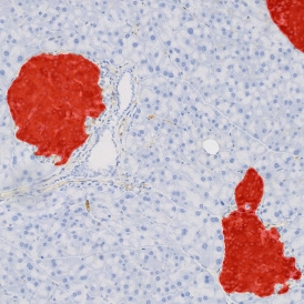
IHC INSULIN ISLETS
The IHC Insulin Islet App detects marker-stained insulin islets, tissue area, and cell phenotypes within and outside the islets. Outputs include tissue and islet area, cell counts, and phenotype distribution.
insulin islets, pancreas, beta-cells
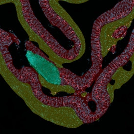
IF SWISS ROLL
The IF Swiss Roll App segments tissue into subclasses (e.g., mucosa, follicles, connective tissue), detects nuclei, and identifies phenotypes via IF stains.
mouse, colon, fluorescence, immune cell follicles
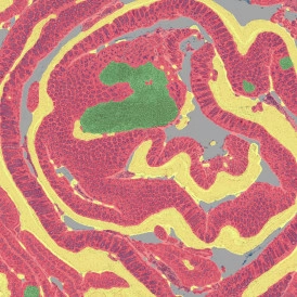
IHC SWISS ROLL
The IHC Swiss Roll App segments the tissue into subclasses (mucosa, follicles, etc.), detects nuclei, and classifies phenotypes. Outputs include tissue areas, cell counts, and phenotype distribution per region.
mouse, colon, immunohistochemistry, immune cell follicles
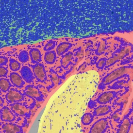
MUCIN SWISS ROLL
The Mucin Swiss Roll App segments tissue into subclasses, detects nuclei and mucin (e.g. PAS-stained), and outputs tissue areas, cell counts, and mucin area per region and overall.
mouse, colon, mucin, immune cell follicles
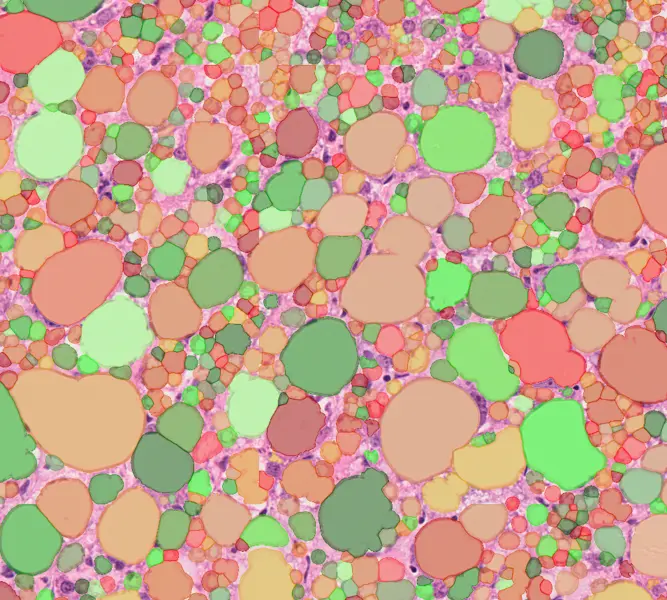
LIPID DROPLETS
The Lipid Droplets App quantifies lipid droplets in H&E-stained tissues, correcting membrane artifacts and providing counts and area measurements.
liver, lipid droplets, H&E
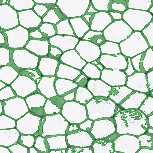
IHC ADIPOCYTE
The IHC Adipocyte App quantifies adipocytes and their lumina in HE samples, correcting membrane artifacts and providing area measurements.
adipocytes, fat tissue, fat cells, H&E
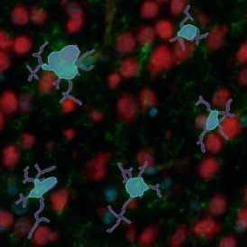
IHC NEURONAL CELLS
The IF Neuronal App detects neuronal cells and branches, quantifies neurites and branching points, and outputs cell area, branch length, and branch counts.
neuronal cells, microglia, central nervous system, peripheral nervous system, phagocytosis, astrocytes, branches, fluorescence
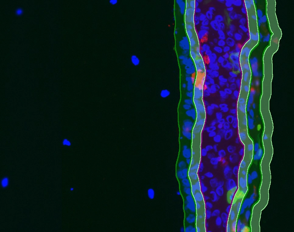
IF ARTIFICIAL SKIN
The IF Artificial Skin App stratifies skin equivalents into dermis and epidermis, further dividing the epidermis into stratum corneum, suprabasal, and basal layers. It outputs area, mean staining intensity, nuclei counts, and % of marker-positive cells for each layer and sublayer. Read publication here
dermatology, epidermis, dermis, skin, aging, oxidative UV damage, artificial skin

