IHC Lung Cancer Mouse
Segment murine lung sections into tumor and non-cancerous tissue using a machine learning classifier, detect hematoxylin-stained nuclei, and quantify tumor area and marker-positive cellular phenotypes.
metastructures
single-cell analysis
tumor microenvironment
immunohistochemistry, lung cancer, tumor microenvironment, mouse, lung
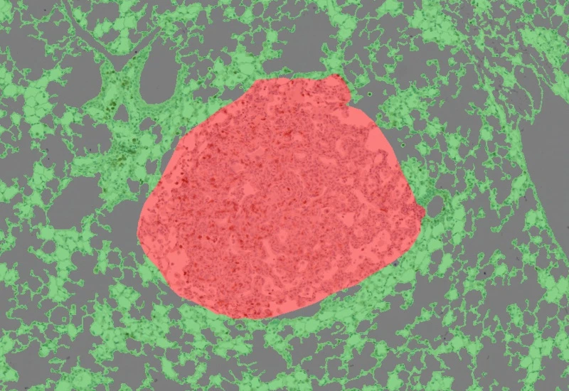
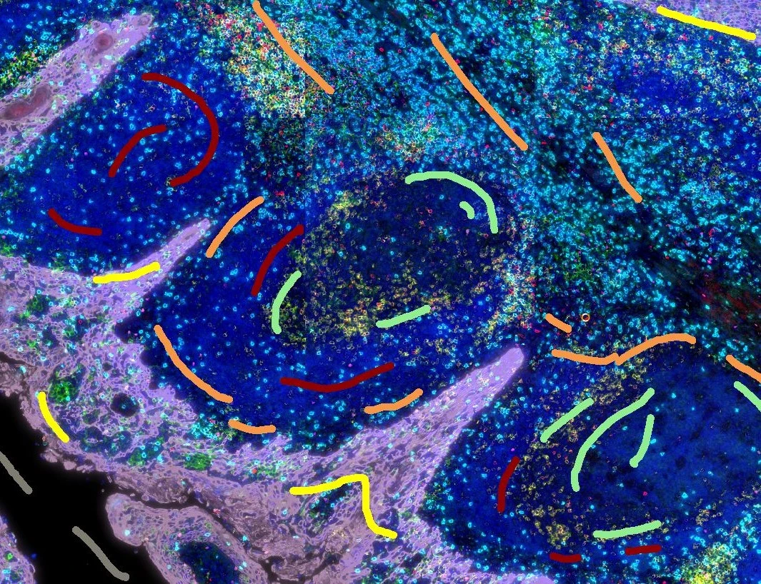
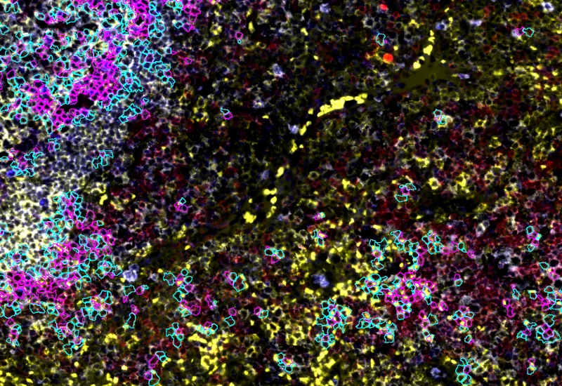
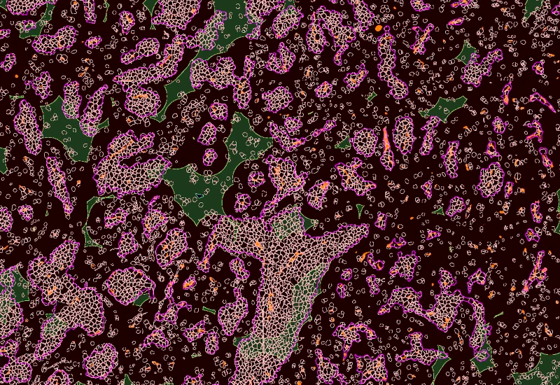
The IHC Lung Cancer mouse App is using the machine learning base classifier to segment murine lung cancer tissue sections into tumor and non-cancerous tissue. Further it detects nuclei based on hematoxylin staining and identifies cellular phenotypes based on speicifc markers. It outputs number and area of tumor tissue as well as total number of cells and detected cellular phenotypes.
Image courtesy of Emma Nye, Head of Experimental Histopathology, The Francis Crick Institute, London, UK
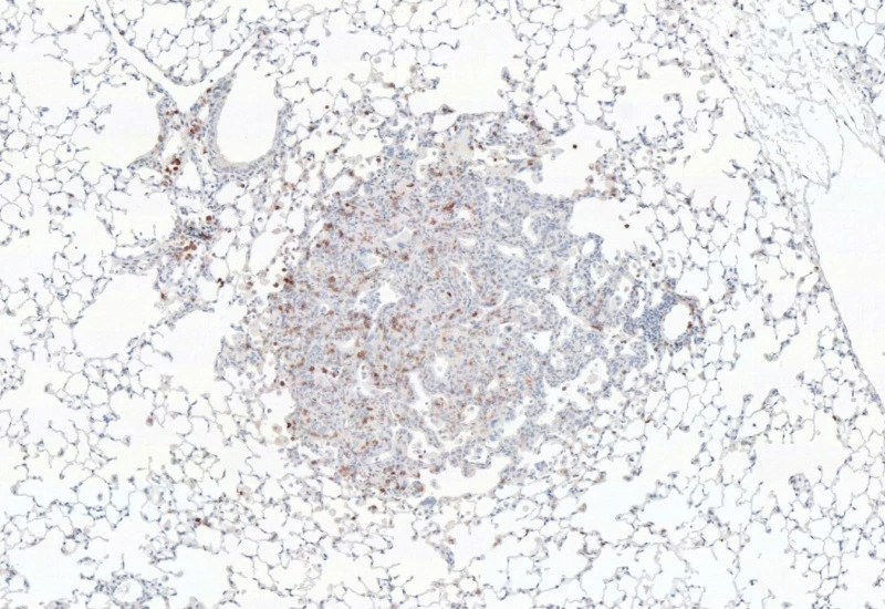
Original Image
Tumor and tissue detection
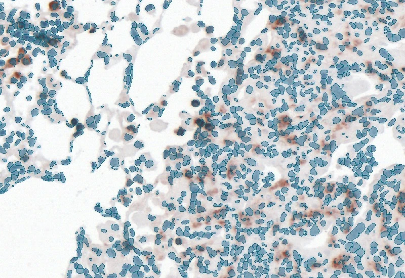
Nuclei detection
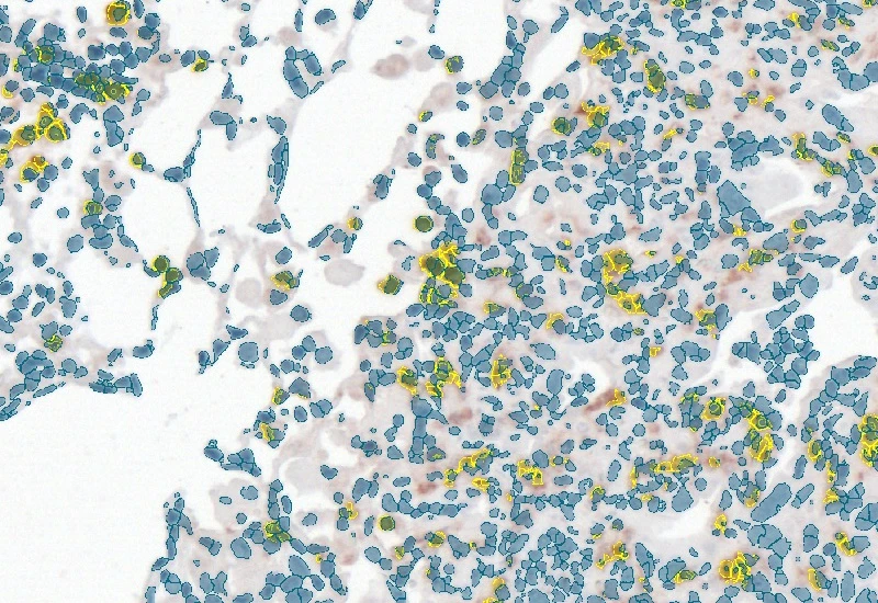
Phenotype detection
tumor microenvironment
Blog Post
07 Apr, 2026
How immunofluorescence image analysis factors into NSCLC studies
single-cell analysis
White Paper
30 Mar, 2026
Understanding NeuroCOVID-19: SARS-CoV-2 Disrupts Astrocyte Homeostatic Functions
metastructures
Blog Post
17 May, 2023
An Intro to Deep Learning in Biomedical Imaging
We support the following file formats:
- TissueFAXS (aqproj)
- StrataFAXS II (vmic)
- PreciPoint (vmic, gtif)
- Generic BigTIFF Import
- Support for multipage BigTIFF files
- OME-TIFF
- JPEG, PNG, BMP, TIFF
- Zeiss (czi)
- Hamamatsu NanoZoomer (ndpi)
- Aperio (svs)
- Leica (scn)
- 3D HISTECH Pannoramic
- Mirax (mrxs)
- Olympus (vsi)
- More slide scanners to be added!
Related Apps
IF Cellular Contact
Determine cellular phenotypes in IF-stained samples, analyze direct cell–cell contacts, and quantify marker intensity, morphometric parameters, and number/% of contacting phenotypes.
cell contacts, spatial interactions, phenotype interactions, B cells, T cells, lung, lymph node, COVID-19, SARS-Cov-2

IHC Angio
Detect blood vessels based on appropriate stains (e.g. CD31), measure vessel and lumen areas, and export vessel number, density, and areas of vessels, endothelium, and lumina.
blood vessels, tumor vascularization, tumor microenvironment, brightfield

Custom App development
Perfectly tailored image analysis solutions for your research.
You have a specific research question that needs to be answered? We offer custom development of image analysis pipelines for specific tasks, be it detection of cellular phenotypes or quantification of tissue structures. After discussing your goals with one of our experts, you will get a ready-to-use App and be a step closer to an impactful publication.