Latest articles in

White Paper
20 Feb, 2019
Imaging of neural contributions to voluntary hand movement
Researchers quantified sensory and motor axons in the human brachial plexus using ChAT and neurofilament immunofluorescence. Automated whole-slide analysis revealed that sensory axons vastly outnumber motor axons, providing new insights into the neural control of fine hand movements.
White Paper
20 Jul, 2016
Digital Image Analysis vs. Pathologist Scoring for ERβ2 Immunohistochemistry in Prostate Cancer
A study in Diagnostic Pathology compared traditional pathologist scoring with digital image analysis for ERβ2 immunohistochemistry in prostate cancer tissue microarrays. Digital analysis demonstrated higher reproducibility and improved quantitative assessment of biomarker expression.

Application Note
14 Oct, 2024
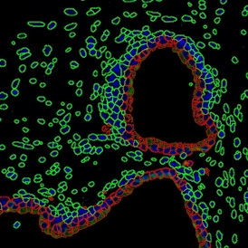
Quantitative Analysis of Cultured Cells
A case study using StrataQuest image analysis on IF-processed cultured cells. The workflow automated cell identification, nuclei and cytoplasm counting, and marker intensity quantification.
White Paper
27 Sep, 2024
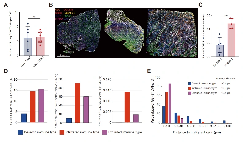
Multimodal Spatial Biology Approach Reveals a Novel Cancer-Associated Fibroblast (CAF) Subset in HNSCC
In HNSCC, CD8+ T-cell infiltration is often impaired. Chuwen Li et al. used mIF, TissueFAXS Spectra, and StrataQuest to image and analyze the spatial distribution of Gal9+ CAFs and CD8+ T cells, complementing prior spatial transcriptomics.
Application Note
14 Feb, 2024
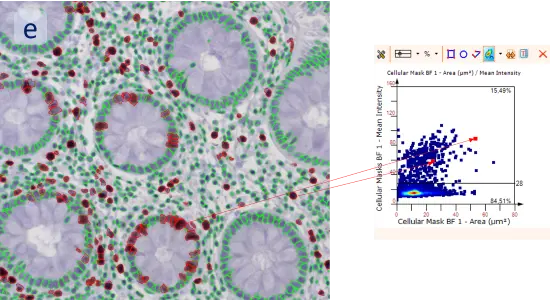
Adipocyte App: Measurement of Cellular Size of Adipocytes
Automated adipocyte detection and size measurement using the StrataQuest Adipocyte App. The workflow identifies adipocytes based on membrane detection and quantifies cell number, diameter, and size in H&E-stained fat tissue.

Webinar
05 Feb, 2024
Unveiling the Dichotomous Role of STAT3 Signaling in Prostate Cancer
Dr. Lukas Kenner presents his research on STAT3’s dual role in prostate cancer. TissueFAXS, HistoQuest, and StrataQuest enabled whole-slide imaging and single-cell quantification.
Application Note
27 Mar, 2023
IHC 2 APP: Ki-67 Nuclear Staining Analysis
This case study demonstrates the use of StrataQuest’s IHC 2 App to quantify Ki-67 nuclear staining in colon tissue. The workflow enabled detection of total cell counts as well as the proportion of proliferating (Ki-67+) cells.
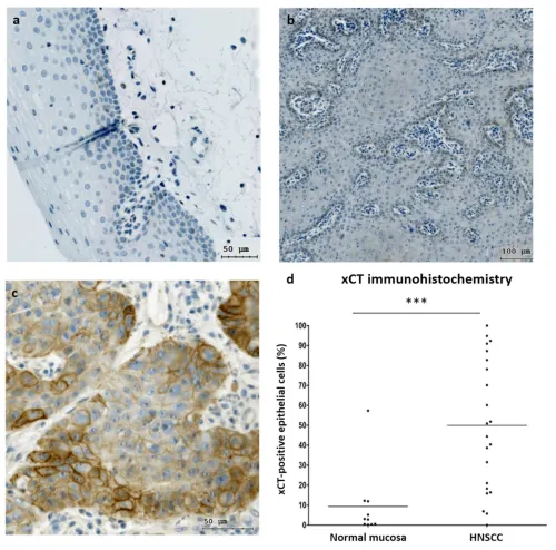
Customer Publication
16 Jan, 2023
Activated Erk 1/2 Kinases Decrease Cell Viability Caused by Erastin
Savic et al. studied the role of Erk 1/2 kinases in ferroptosis induction in HNSCC. They found erastin to be a potent ferroptosis inducer, strongly dependent on Erk 1/2 activity. IHC-stained samples were quantified using TissueFAXS and HistoQuest.
Blog Post
19 Oct, 2022
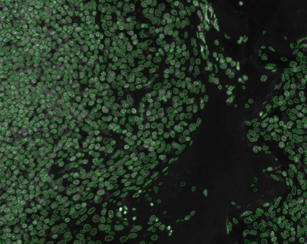
How Deep Learning is Used for Cell Counting
Deep learning is transforming cell counting by enabling precise nuclei segmentation, even in dense tissue microenvironments. Learn how TissueGnostics applies deep neural networks (DNNs) to automate and improve accuracy in tissue cytometry workflows.
Blog Post
12 Oct, 2022
How Cell Counting Algorithms Work
Cell counting is central to biomedical research, but accuracy depends on advanced algorithms. This blog explores manual vs automated methods, and how TissueGnostics’ AI-driven cell counting algorithms enable reliable, high-throughput image-based analysis.
Customer Publication
14 Sep, 2022
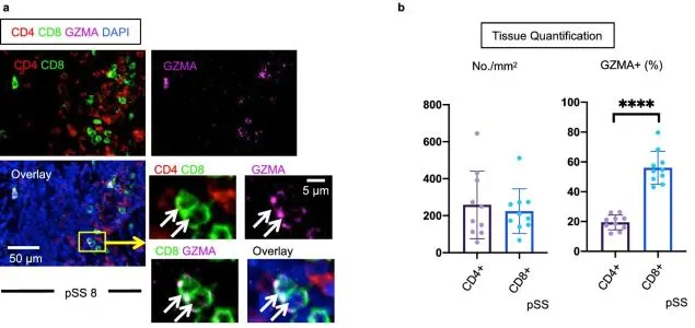
Cytotoxic CD8+ T cell Role in Tissue Destruction in Sjögren’s Syndrome
Kaneko et al. (Scientific Reports, Nature) investigated CD4+ and CD8+ T-cell subsets in salivary glands of Sjögren’s syndrome patients using TissueFAXS with TissueQuest and StrataQuest for in-depth quantitative tissue cytometry.
Customer Publication
06 Dec, 2021
Tissue Cytometry in Anti-SARS-CoV-2 Drug Discovery
Zhang et al. (Signal Transduction Targeted Therapy) studied Azvudine in SARS-CoV-2 infected macaques and COVID-19 patients. TissueFAXS Q and TissueQuest quantified viral load, immune status, and TUNEL staining in thymus tissue.
Blog Post
09 Nov, 2021
What is Quantitative Histopathology?
Histopathology is the scientific branch focusing on assessment of diseases by examining stained biological tissue sections, particularly in human subjects. This field can give immense insight into biological and medical areas of study.