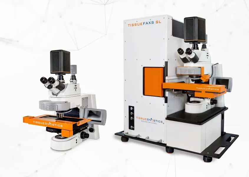
TissueFAXS
Multispectral Imaging Systems
Multispectral whole-slide imaging for increased marker detection and cleaner signal.
See what’s new in Version 8 ›
Software Suite 8 is here — DNN tissue detection, label capture with existing cameras, and a redesigned interface with smarter, more intuitive tools.
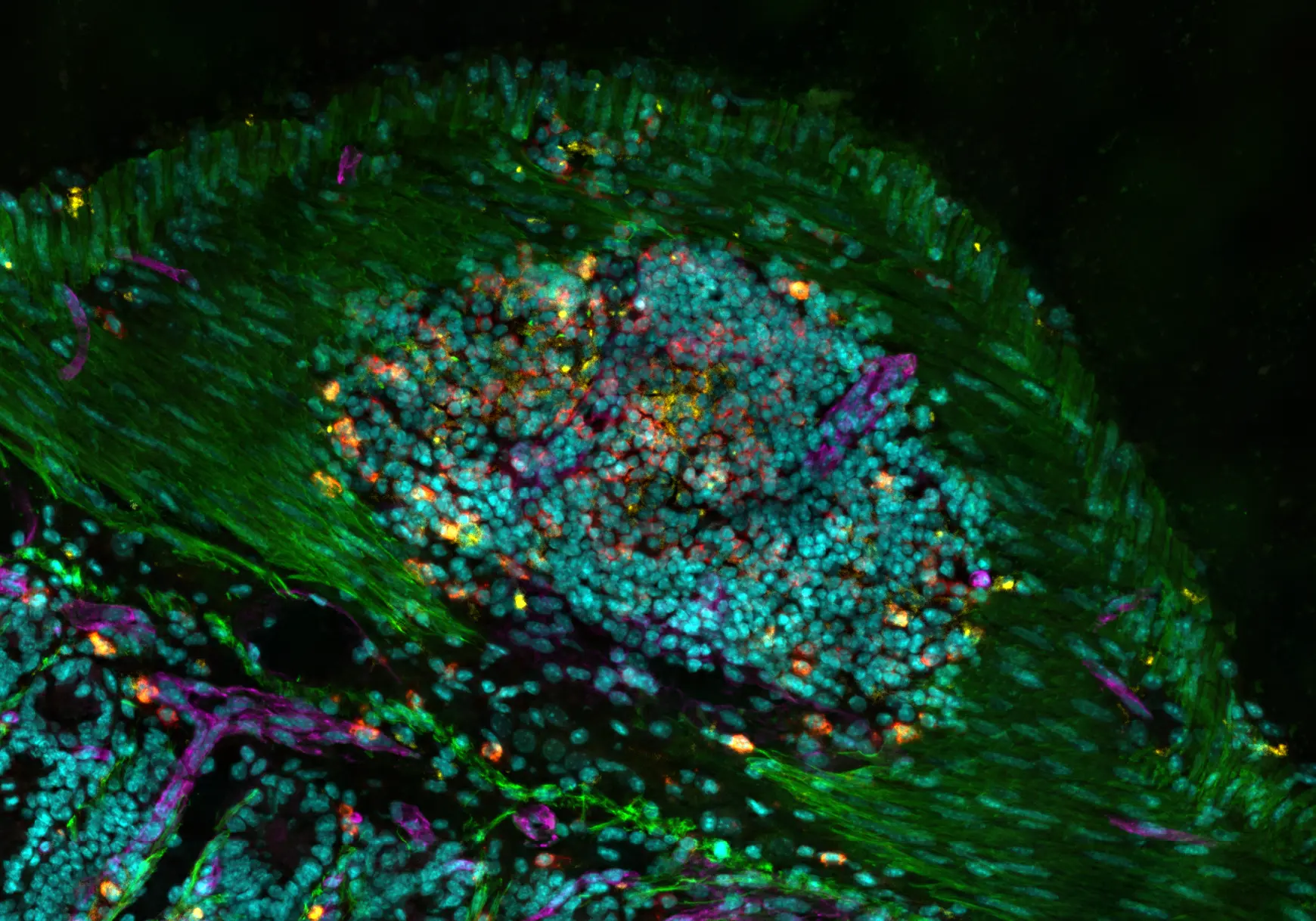
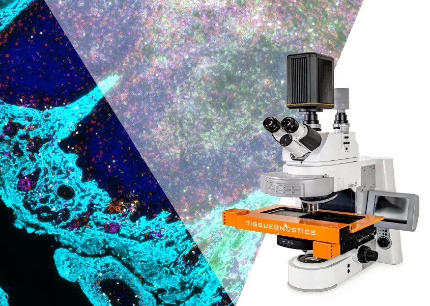

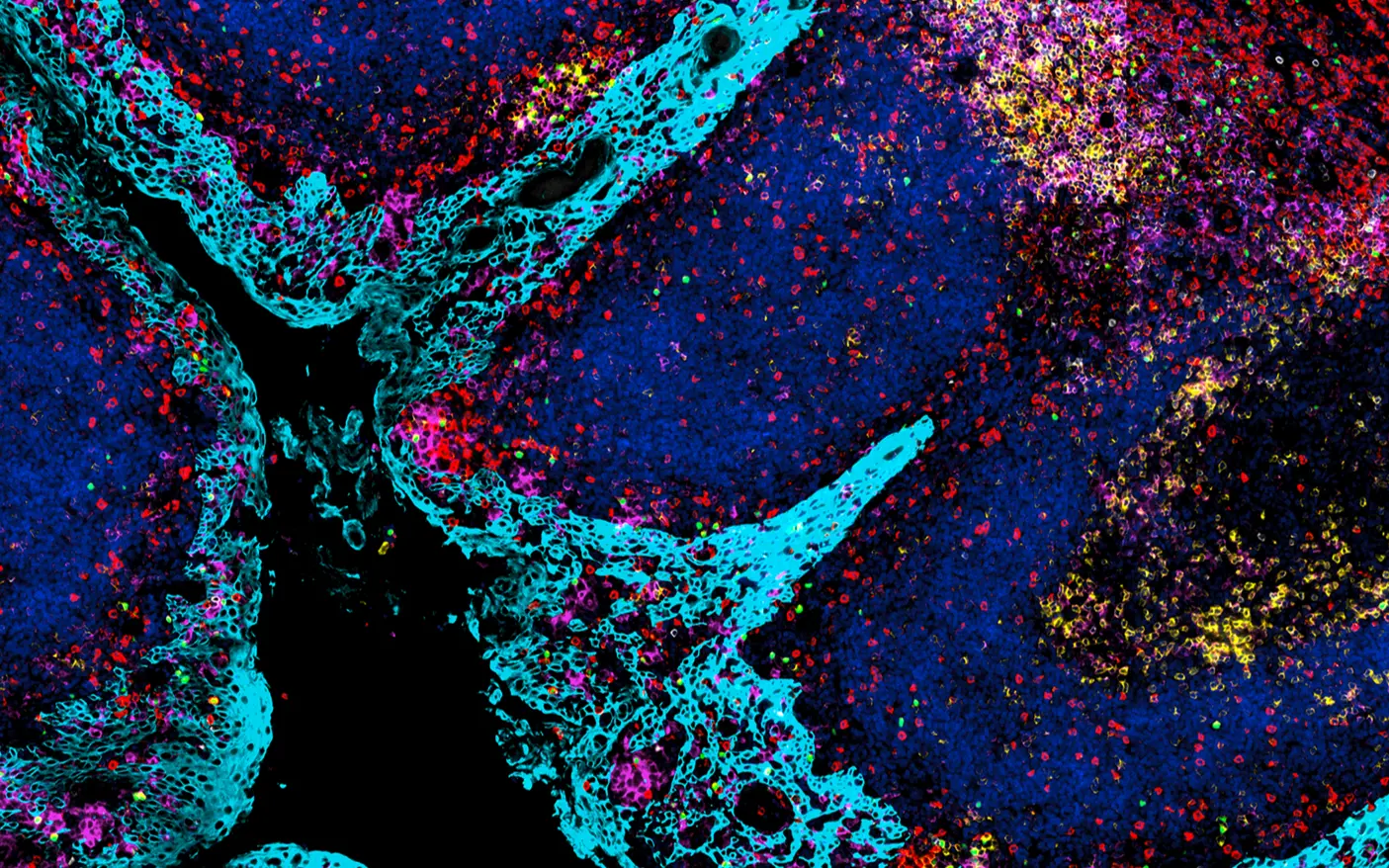
Expand the Limits of Multiplex Imaging
High-content fluorescence imaging for deeper insights.
The TissueFAXS Spectra and Spectra SL systems are designed for seamless multispectral whole-slide imaging. Using liquid crystal tunable filter (LCTF) technology, they allow in-situ phenotyping of up to 8 markers per scan with minimal channel bleed-through and optimized spectral separation. These systems support complex research demands like immune profiling or spatial biology, with downstream analysis tools ensuring ready-to-publish results.
- Up to 8-marker multiplexing per scan
- Spectral unmixing to reduce autofluorescence and channel bleed-through
- Extensive Reference Spectra Database for spectral unmixing
- Quantify phenotypes and spatial patterns
- Upgrade-ready for 120 slide automation
See More with TissueFAXS Spectra
Our multiplex imaging platform delivers unmatched insights into the cellular composition of your sample
See More in One Scan
Capture up to 8 fluorescent markers in a single run - perfect for complex tissue characterization.
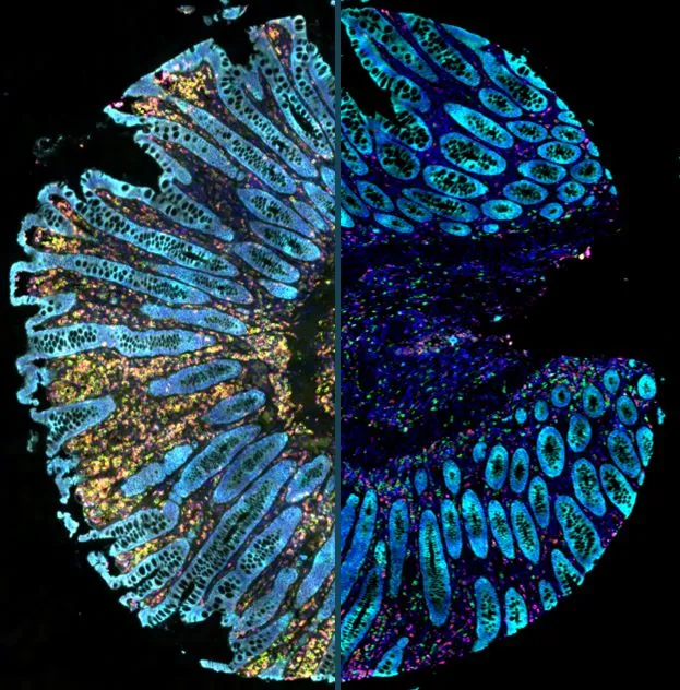
Pure Signal, No Noise
Remove autofluorescence and channel bleed-through with spectral unmixing for accurate signal detection.
Smart Integrated Analysis
Transfer scans directly to analysis and identify and quantify phenotypes with ease.
Expert support you can count on
From setup to publication, our team helps you maximize multispectral imaging performance.
Experience Signal Clarity Like Never Before.
Visualize every detail with confidence - TissueFAXS Spectra’s advanced optics and spectral unmixing ensure crisp, artifact-free imaging


Key features of Spectra Systems
- Expanded Multiplex Imaging Capabilities
- Spectral Unmixing
- All-in-one Workflow
- Open Assay Design
- Powerful Imaging Software
- Modular and Scalable Design
- Walkaway Automation
- Component Flexibility
- Integrated Image Analysis
- Unmatched Expert Support

Expanded Multiplex Imaging Capabilities
Using liquid crystal tunable filter (LCTF) technology, the system is capable of detecting more markers in a single scan, enabling detailed visualization of complex tissues while saving time and resources. The ability to tune the filter permits for capturing a wide range of wavelengths, making it a highly flexible tool for multiplex fluorescence imaging.


Extensive Reference Spectra Database and Spectral Unmixing
TissueFAXS Spectra provides a large database that characterizes the unique emission spectra of various commonly used fluorochromes. Those can be used for spectral unmixing of the scanned image, and as a result, potential channel bleed-through as well as autofluorescence is eliminated, delivering a clear image.
All-in-one Workflow
Our platform offers a streamlined, step-by-step workflow that simplifies the entire process, from multispectral whole-slide imaging and spectral unmixing to delivering a digital high-quality view of the native tissue environment with seamless stitching.

Open Assay Design
No need to change existing staining workflows - with our system you can use your own antibodies. TissueFAXS Spectral is compatible with many commercially available staining kits, such as Opal or Alexa Fluor Dyes - reach out to our team for specifications.


Powerful Imaging Software
TissueFAXS features powerful imaging software designed to support both routine and advanced workflows. Users have full access to all scanning parameters allowing protocols to be precisely tailored and saved as reusable profiles for consistent results. Smart autofocus and Z-stacking with a Z-projection (extended focus) algorithm, improve clarity in thick or uneven samples, resulting in high-quality seamless images.
Integrated virtual slide viewing, annotation tools, and export to open data formats streamline review, sharing, and downstream analysis.

Modular and Scalable Design
TissueFAXS Multispectral is designed to perfectly suit your research needs - it comes equipped with an 8-slide stage ideal for a smaller capacity lab. Need to scan H&E or IHC-stained tissues too? TissueFAXS Multispectral supports both fluorescence and brightfield imaging modes. Plan a high-throughput research project? A 120-slide loader can be added for high-throughput studies or core facilities. Thanks to the system flexibility, your TissueFAXS system can grow with your lab.

Walkaway Automation and Integrated Label Reader
TissueFAXS supports walk-away automation with a fully motorized, computer-controlled system and automated workflows for a wide range of sample types and staining protocols. Key steps - like tissue detection, autofocus, and image acquisition - are handled automatically for both brightfield and fluorescence experiments.
Built-in barcode and QR code reading captures sample IDs during scanning, allowing for automatic slide tracking and accurate metadata assignment. With customizable scan profiles and batch processing, TissueFAXS makes it easy to run multi-slide projects unattended - while ensuring traceable, consistent, and reproducible results.

Component-Flexibility (Objectives, Cameras, Light Sources, Filters)
Users can choose from a variety of high-performance cameras optimized for both brightfield and fluorescence imaging; objectives ranging from 1x to 100x in air, water, or oil immersion - with support for up to seven simultaneously mounted; multiple light sources with configurable spectral outputs to match specific fluorophore combinations; and filter sets including single-, dual-, and quad-bandpass options.
This component-level flexibility allows configurations to be precisely tailored to specific experimental needs, from routine imaging to advanced multiplexed assays.

Seamless Integration with Image Analysis
The Multispectral configuration is built for high-plex imaging - and StrataQuest provides the tools to turn that complexity into clarity. Fully integrated spectral unmixing feeds into advanced analysis workflows like marker co-expression, multiplex phenotyping, and neighborhood analysis. These allow for quantifying several markers per cell and extracting spatial patterns in the tumor microenvironment, lymphoid structures, or autoimmune lesions. Whether resolving 4 or 40 markers, the imaging and analysis workflow remains streamlined, accurate, and ready to scale with your project.

Specialized Expertise, Every Step of the Way
Our systems are backed by a multidisciplinary support team with expertise across hardware configuration, image analysis, system maintenance, and biology.
Our application specialists help you define the optimal setup for your imaging needs, while our analysis experts assist in designing and refining StrataQuest pipelines tailored to your research. For more advanced needs, engineering and R&D teams are available to develop and integrate custom features or workflows - including new configurations beyond our standard portfolio.
Whether you're scaling your analysis or planning for future upgrades, expert guidance is built into every step.
High-Throughput Multispectral Whole-Slide Imaging
TissueFAXS Spectra SL
Replace the standard 8-slide stage with a high-capacity automated loader - instantly transforming your system into a high-throughput imaging platform.
- Combines multispectral imaging with 120-slide batch scanning.
- Supports mixed sample types and imaging modes in a single unattended run.
- Ideal for high-content phenotyping, spatial analysis, and large studies.
- Built for labs handling multiplexed assays at scale - without sacrificing image quality.
Interested in seeing TissueFAXS Spectra in action?
Explore TissueFAXS Spectra in Action
Our knowledge base provides valuable resources where StrataQuest was employed to push the science forward - white papers, application notes and webinars.

Multimodal Spatial Biology Approach Reveals a Novel Cancer-Associated Fibroblast (CAF) Subset in HNSCC

Spectral Imaging and Spatial Analysis of TMA Samples
Analysis of organoid and immune cell co-cultures with machine- learning-powered image analysis
Related Products

TissueFAXS Viewer
Free. Fast. Made for Collaboration.
TissueFAXS Viewer is a free, standalone desktop application for exploring high-resolution virtual slides generated by TissueFAXS systems. It allows researchers and collaborators to navigate brightfield, fluorescence, or multispectral scans, view annotations, and assess results without needing the full imaging setup. It's the ideal tool for sharing and reviewing data across teams.
StrataQuest Image Analysis
Turns imaging data into publication-ready results.
StrataQuest is a powerful analysis platform designed to work seamlessly with TissueFAXS. It transforms high-resolution images into meaningful data, supporting workflows from basic segmentation to AI-driven classification. Scalable and intuitive, it delivers insights into phenotypes, spatial patterns, and tissue architecture across a range of research applications.
- Customizable pipelines for basic & advanced image analysis
- AI-powered nuclei segmentation and tissue classification
- Spatial analysis tools (e.g. neighborhood, distance, clustering)
- Integrated data mining: t-SNE, UMAP, SONG, and more
- Prebuilt analysis apps for oncology, immunology, neurology, and more
- Open format support and direct integration with TissueFAXS systems

Let Us Know How We Can Support You
Couldn't find the answer to your question? Need assistance?
Whether you're imaging slides, well plates, or complex 3D samples, our team is here to help configure a TissueFAXS system that fits your exact workflow. From hardware recommendations to future upgrade planning, we’ll work with you to design a setup that’s ready for now and built for what’s next.
Technical Specifications
Feature | TissueFAXS SPECTRA - 8 Slide Stage | TissueFAXS SPECTRA SL - High-Throughput | ||
|---|---|---|---|---|
Sample Carrier | Slide Types | glass slides | ||
Compatible Glass Slide Formats | Standard slide: 25 mm x 75 mm (1 in × 3 in) | Standard slide: 25 mm x75 mm (1 in × 3 in) | ||
Thickness Cover Glass | 0.12 mm - 0.17 mm | |||
Design | Imaging Modes | multispectral imaging using LCTF filter, widefield fluorescence, brightfield*, darkfield*, phase contrast*, polarization*, DIC* | ||
Capacity | 8 standard slides | 120 standard slides or 60 double sized slides | ||
Objectives | motorized nosepiece for up to 7 (1x - 100x) air, oil or water | motorized nosepiece for up to 6 (1x - 100x) air oil or water | ||
Stage | motorized 8 slide stage (x,y,z) | automated slide loader for up to 120 slides | ||
Workstation | HP Z6, High-performance Windows 11 PC for microscope control and analysis, including 2 x 27 high quality TFT monitors | |||
Brightfield* | Light Source | VIS-LED: Lifetime > 10.000 hours | ||
Color Camera | Pixelink (CMOS): 2048 x 2048 px; 90.3 fps; up to 63% QE | |||
Scan Time | approx. 1.5 min (15 x 15 mm, 20x objective, pure scanning time, 1 focus point) | |||
Fluorescence | Light Source | Spectra III: 8 solid-state LEDs operating independently; 390-680 nm; ~500 mW power output/LED | ||
Monochrome Camera | Options: | |||
FL-Channels | motorized filter wheel, up to 10 positions | |||
Scan Time | Standard FL scan: approx. 9 min (15 x 15 mm, 20x objective, 4 channels, 10ms exposure time, pure scanning time, 1 focus point) | |||
Dimensions | Measurements | 800 mm x 700 mm x 750 mm (31.5 in x 28 in x 30 in) | 750 mm x 1250 mm x 1404 mm (29.5 in x 50 in x 55 in) | |
Weight | 80 kg ± 5% (176.4 lbs ± 5%) | 184 kg ± 5% (405.7 lbs ± 5%) | ||
Operation | Temperature | +10 to +30°C (50 to 86°F) | ||
Humidity | below 40% is recommended; up to 80% without condensation | |||
Power Consumption | minimum 500 watts at peak, minimum of two power sockets 110/220 Volts (US/Europe) | |||
Software | Scanning | spectral unmixing, reference spectral database, one click workflow, customizable workflows, access to all scanning parameters, automatic AI based tissue detection, automatic label scanning, HDR and deconvolution**, smart autofocus, z-stacks and z-projections, automatic stitching for seamless whole slide image, dedicated TMA and well-plate workflow | ||
Image Export | aqproj (bioformats), OME tiff, tiff, png, jpg, bmp | |||
Image Viewer | Free TissueFAXS viewer - download here | |||
Analysis* | AI-based nuclei segmentation, real-time analysis preview, phenotyping and phenotype interactions, proximity measurement tool, AI training center**, dot detection, interactive diagrams, omics explorer**, unlimited multiplexing, library of prebuilt pipelines, data mining tools, customized statistics, versatile data export | |||
Trusted by Thousands of Researchers
Over 3,200 publications using TG solutions
Researchers worldwide rely on TG imaging and analysis platforms to generate high-quality, reproducible data across a wide range of fields—from oncology and immunology to neuroscience and regenerative medicine.
