Quantification of p-H2AX Foci in Co-cultured Cells Exposed to Radiation, Livia Sima
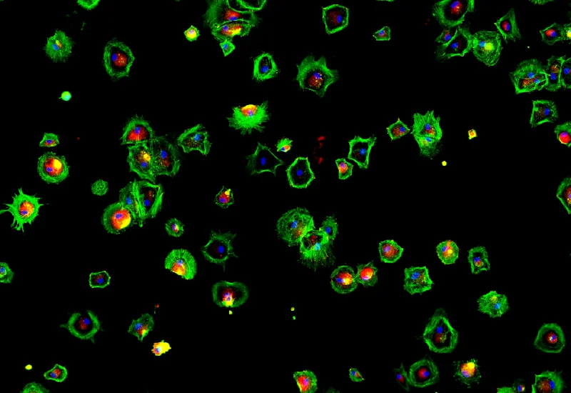
Dr. Livia Sima, Romanian Academy, presents FLASH radiation therapy and its effect on melanoma cells. The team used TissueFAXS iPLUS slide scanner for imaging the cells and TissueQuest for image analysis.
Webinar
Quantification of p-H2AX Foci in Co-cultured Cells Exposed to Radiation, Livia Sima
19 Nov, 2024
Dr. Livia Sima, Romanian Academy, presents FLASH radiation therapy with short-pulse high-dose delivery and its effect on melanoma cells. The results showed its effectiveness against cancer cells, whereas normal cells are not affected by the treatment. The team used TissueFAXS iPLUS slide scanner for imaging the cells and TissueQuest for image analysis.
Key Takeaways
- Quantifying DNA damage with imaging: Learn how γH2AX foci can be used as a reliable marker to measure DNA double-strand breaks in cells exposed to radiation.
- Automated single-cell analysis: Discover how TissueFAXS and TissueQuest enable high-throughput, single-cell quantification of nuclear foci across thousands of cells.
- Practical analysis workflow: The webinar demonstrates a full analysis pipeline, from slide scanning and nuclei segmentation to gating strategies and statistical data export.
- Comparing radiation modalities: Results illustrate how FLASH radiation may induce different DNA damage responses in melanoma cells versus normal melanocytes.
- Advanced research applications: The approach supports studies on radiation therapy, DNA damage response, and differential cellular sensitivity in cancer research.