
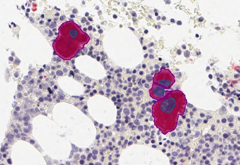
IF Cell Culture - Osteoclast
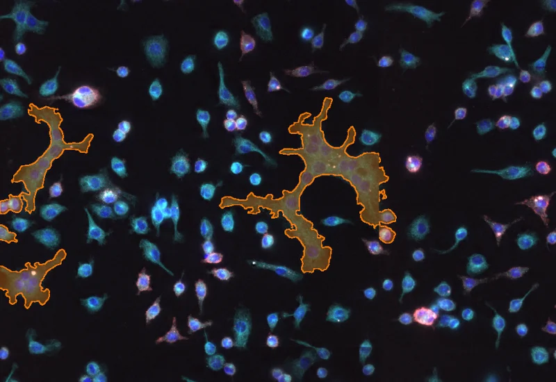
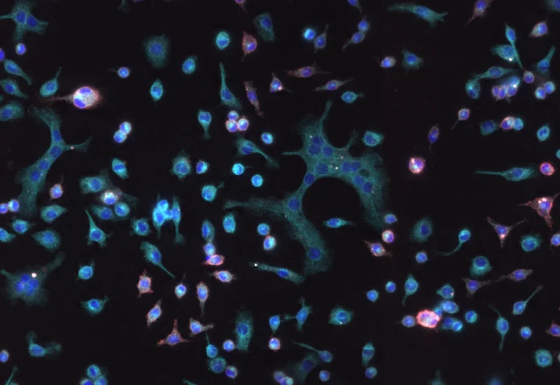
Segment nuclei, identify multinucleated osteoclasts, and quantify cell number, nuclei per osteoclast, osteoclast area, and marker intensity within osteoclasts.
bone research, osteoclast, multinucleated cells, bone adsoprtion
The IF Cell Culture - Osteoclast App allows segmentation of nuclei, the identification of cultured multinucleated osteoclasts (stained by a specific marker) and if wanted the quantification of one or two additonal markers. The App outputs the number of detected cells, osteoclasts and nuclei per osteoclast as well as the area of osteoclasts and intensity of markers within the osteoclasts.
Heindl, A. et al. (2016). Toward the Automated Detection and Characterization of Osteoclasts in Microscopic Images. In: Pietschmann, P. (eds) Principles of Osteoimmunology. Springer, Cham. https://doi.org/10.1007/978-3-319-34238-2_2
Prof. Peter Pietschmann & Isabella Ellinger
Original Image
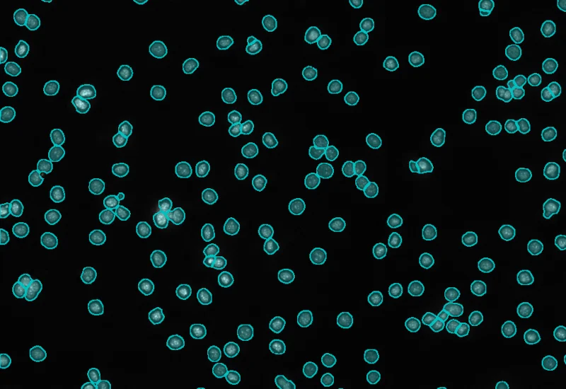
Nuclei detection
Osteoclast detection

Application Note
14 Oct, 2024
Quantitative Analysis of Cultured Cells

White Paper
17 Jul, 2024
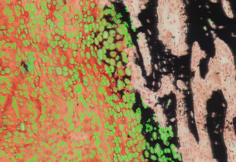
Algorithmic automation of bone research with StrataQuest
White Paper
17 Oct, 2025
Integrative Multiomics Approach Unveils Systemic Dysfunction in Colorectal Cancer (CRC)
We support the following file formats:
- TissueFAXS (aqproj)
- StrataFAXS II (vmic)
- PreciPoint (vmic, gtif)
- Generic BigTIFF Import
- Support for multipage BigTIFF files
- OME-TIFF
- JPEG, PNG, BMP, TIFF
- Zeiss (czi)
- Hamamatsu NanoZoomer (ndpi)
- Aperio (svs)
- Leica (scn)
- 3D HISTECH Pannoramic
- Mirax (mrxs)
- Olympus (vsi)
- More slide scanners to be added!
Related Apps
IHC Megakaryocytes
Detect megakaryocytes in IHC-stained sections, reconstruct full cell area, and quantify cell size, number of megakaryocytes containing neutrophils, and neutrophil count per megakaryocyte.
bone tissue, bone marrow, megakaryocytes, immunohistochemistry

Custom App development
Perfectly tailored image analysis solutions for your research.
You have a specific research question that needs to be answered? We offer custom development of image analysis pipelines for specific tasks, be it detection of cellular phenotypes or quantification of tissue structures. After discussing your goals with one of our experts, you will get a ready-to-use App and be a step closer to an impactful publication.
