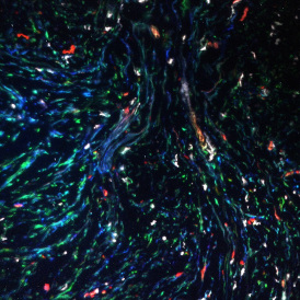
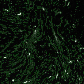
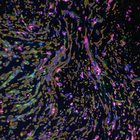
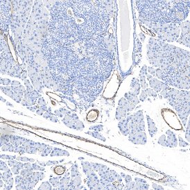
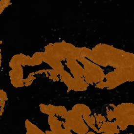
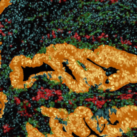
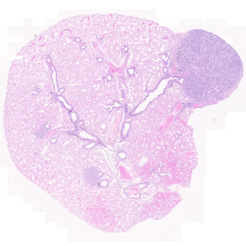
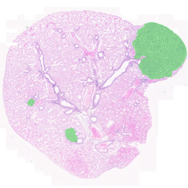
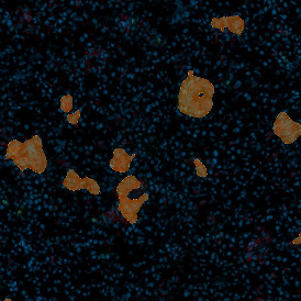
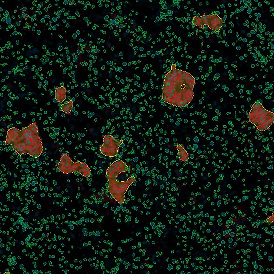
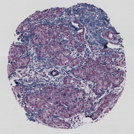
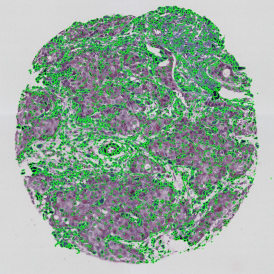
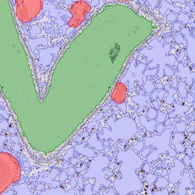
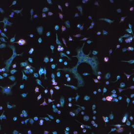
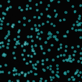
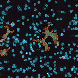
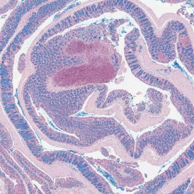
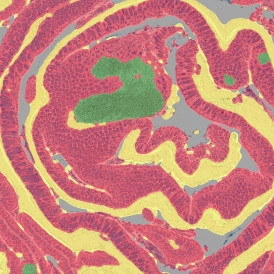
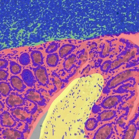
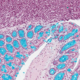
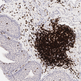
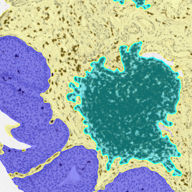
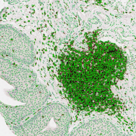
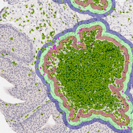
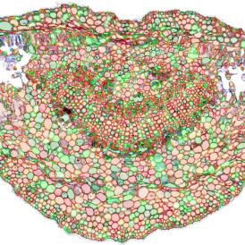
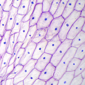
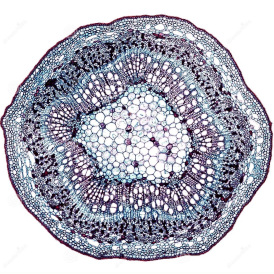
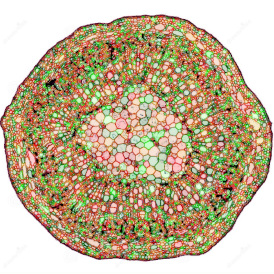
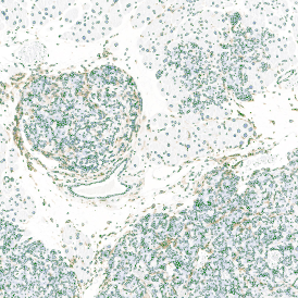
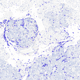
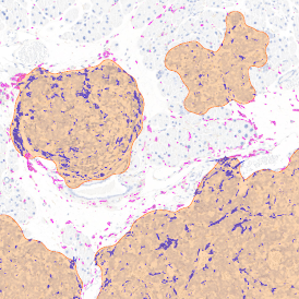
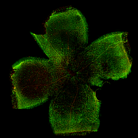
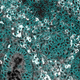
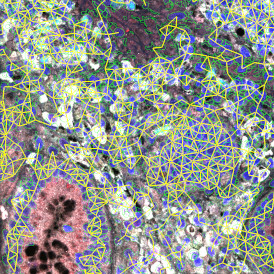
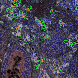
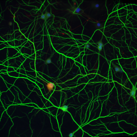
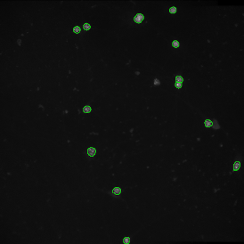
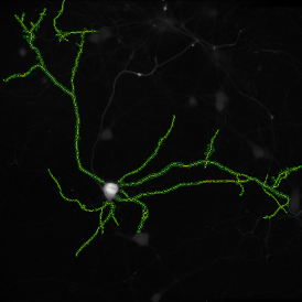
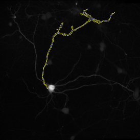
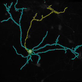
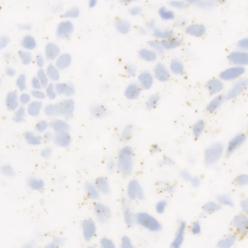
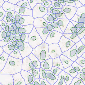
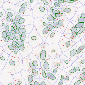
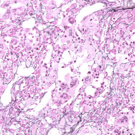
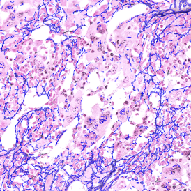
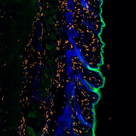
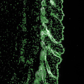
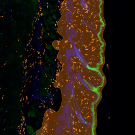
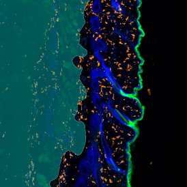
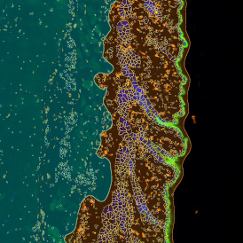
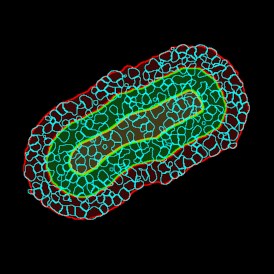
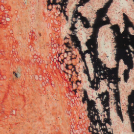
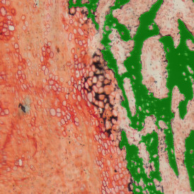
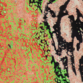
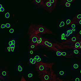
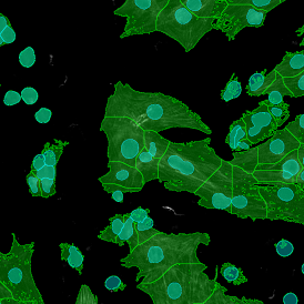
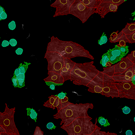
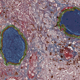
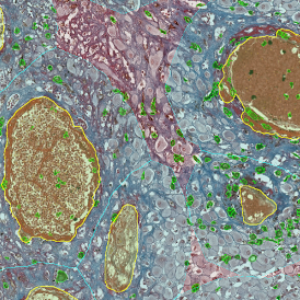
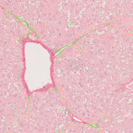
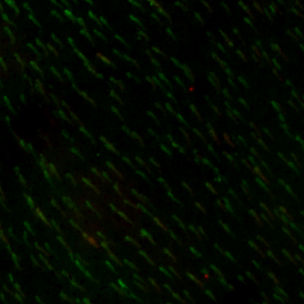
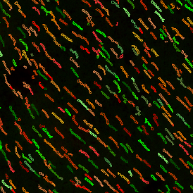
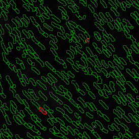
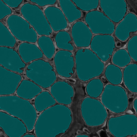
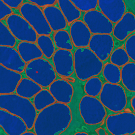
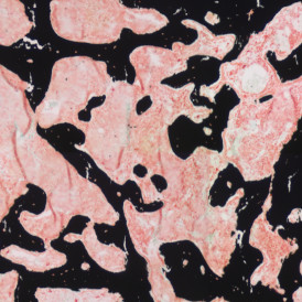
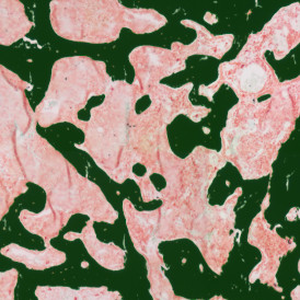
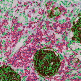
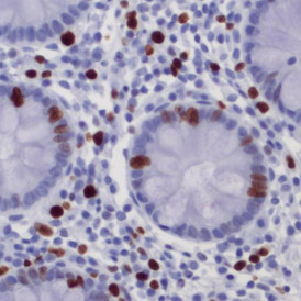
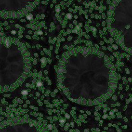
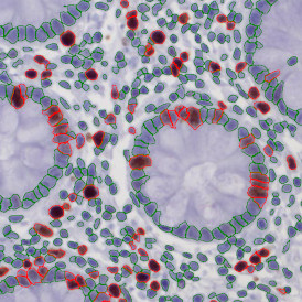
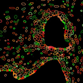
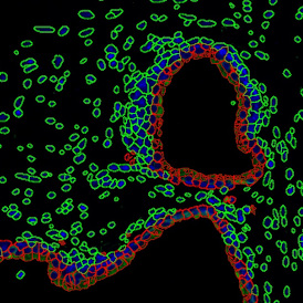
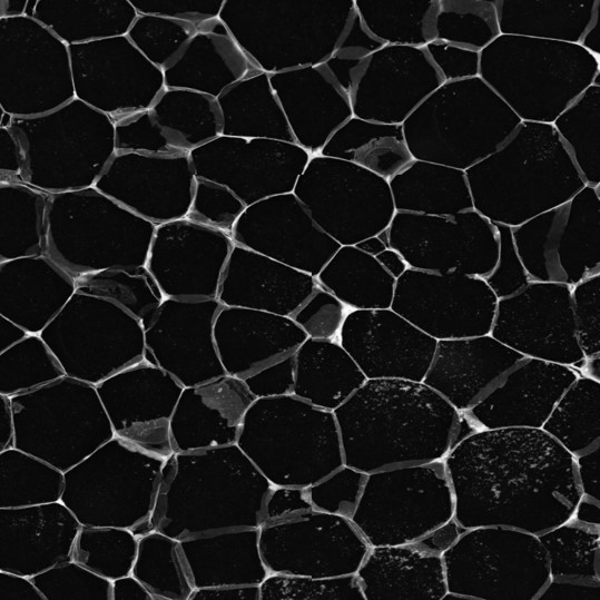
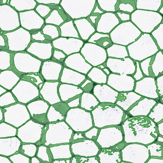
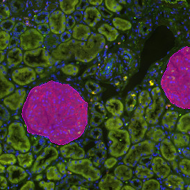
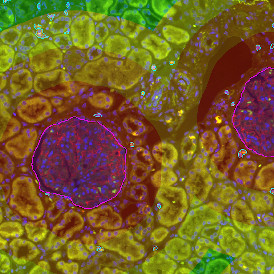
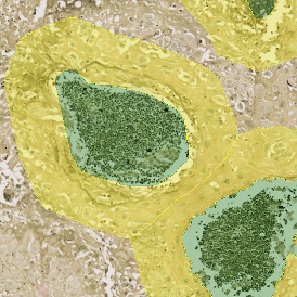
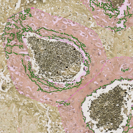
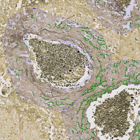
Welcome to the App Center

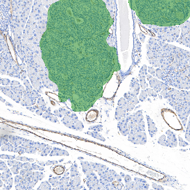
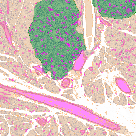
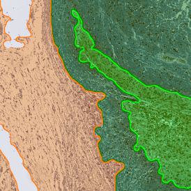
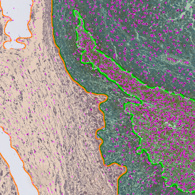
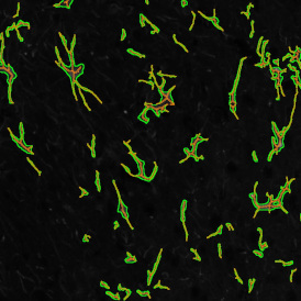
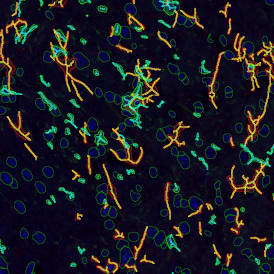
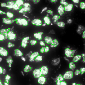
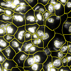
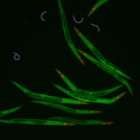
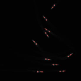
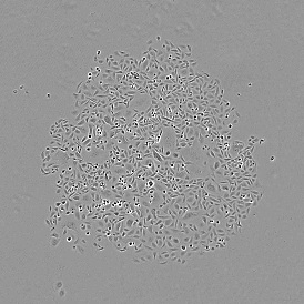
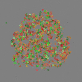
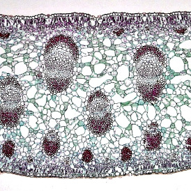
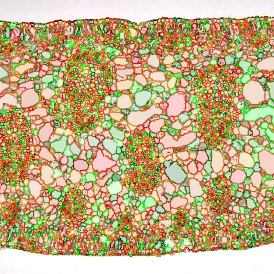
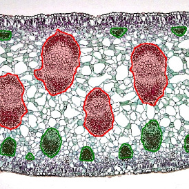
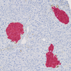
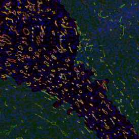
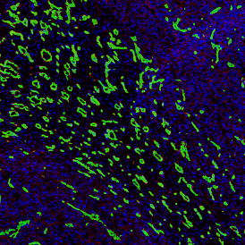
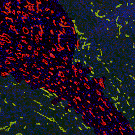
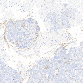
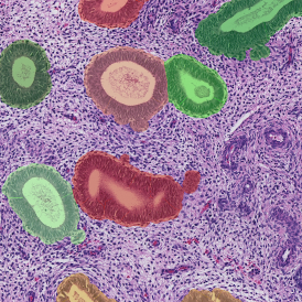
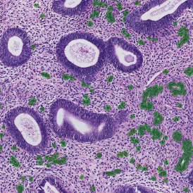
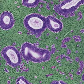
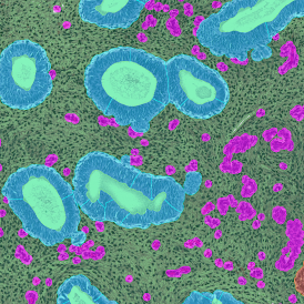
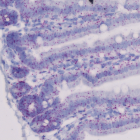
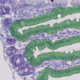
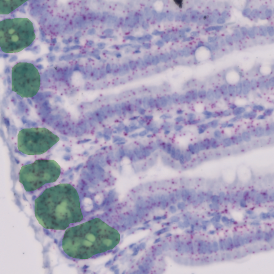
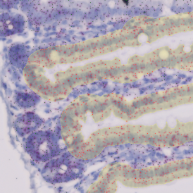
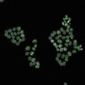
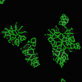
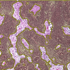
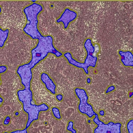
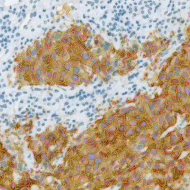
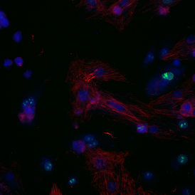
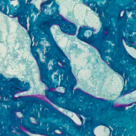
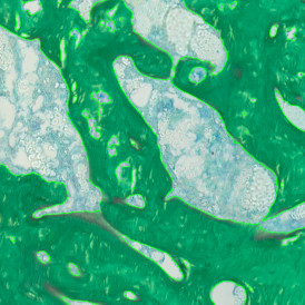
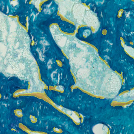
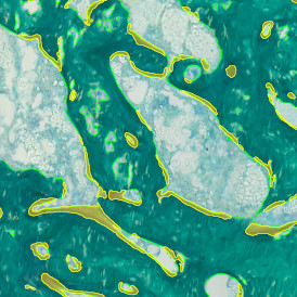
Look through our StrataQuest Apps and get inspired by the variety of biomedical image analysis applications. If you cannot find the perfect solution for your research question please contact us. TissueGnostics offers the development of customized Apps for your specific analysis needs. Contact us for pricing details.

Original image
Nuclei detection
Marker detection
Hi-Plex marker detection
previous arrowprevious arrow
next arrownext arrow
 
IF HI-PLEX 50
Original image
Tumor detection
Vessel detection
Combined detection
previous arrowprevious arrow
next arrownext arrow
 
IHC TUMOR VASCULARIZATION
Original image
Nuclei detection
Tumor detection
Proximity measurements: 0-25µm
Proximity measurements: 25-50µm
previous arrowprevious arrow
next arrownext arrow
 
IF IMMUNE STATUS IN SITU
Original image
Tumor foci detection
previous arrowprevious arrow
next arrownext arrow
 
TUMOR FOCI
Original image
Nuclei detection
Granuloma detection
Combined mask
previous arrowprevious arrow
next arrownext arrow
 
IF GRANULOMA
Original image
Nuclei detection
Tumor detection
Tumor cell detection
previous arrowprevious arrow
next arrownext arrow
 
IHC TUMOR-STROMA
Original Image
Marker-positive cell detection
Stroma/Tumor/Necrotic tissue detection
Combined detection
previous arrowprevious arrow
next arrownext arrow
 
IHC NECROTIC TUMOR
Original image
Glial cell detection
Combined mask
previous arrowprevious arrow
next arrownext arrow
 
IF GLIAL CELLS
Original image
Nuclei detection
Cytoskeleton detection
Dot detection
Cell detection
previous arrowprevious arrow
next arrownext arrow
 
IF CYTOSKELETON
Original image
Nuclei and marker detection
Dermis/Epidermis detection
Combined mask
previous arrowprevious arrow
next arrownext arrow
 
IHC MACROPHAGES
Original image
Nuclei detection
Bronchiole detection
Aveoli detection
Blood vessel detection
Combined mask
previous arrowprevious arrow
next arrownext arrow
 
PULMO
Original Image
Nuclei/cell detection
Red dot detection
Green dot detection
previous arrowprevious arrow
next arrownext arrow
 
RNA SCOPE+
Original image
Nuclei detection
Cell detection
Parasite detection
Combined mask
previous arrowprevious arrow
next arrownext arrow
 
IF LEISHMANIASIS
Original Image
Vessel and vessel surrounding detection
Elastin detection
Collagen detection
previous arrowprevious arrow
next arrownext arrow
 
IHC ANGIO ELASTIN
Original image
Nuclei detection
Glomeruli detection
Proximity measurements
previous arrowprevious arrow
next arrownext arrow
 
IF GLOMERULI
Original Image
Color separation
Membrane detection
Adipocyte detection
previous arrowprevious arrow
next arrownext arrow
 
IHC ADIPOCYTE
Original image
Nuclei detection
Cellular mask detection
Marker-positive cell detection
previous arrowprevious arrow
next arrownext arrow
 
IF 2
Original image
Nuclei detection
Cellular mask detection
Marker-positive cell detection
previous arrowprevious arrow
next arrownext arrow
 
IF 3
Original image
Nuclei detection
Cellular mask detection
Marker-positive cell detection
previous arrowprevious arrow
next arrownext arrow
 
IF 4
Original image
Nuclei detection
Marker-positive cells
previous arrowprevious arrow
next arrownext arrow
 
IHC 2
Original image
Nuclei detection
Marker 1-positive cells
Marker 2-positive cells
previous arrowprevious arrow
next arrownext arrow
 
IHC 3
Original image
Bone detection
Bone marrow detection
Bone surface detection
previous arrowprevious arrow
next arrownext arrow
 
BONE VON KOSSA
Original Image
Lumen detection
Myofibre detection
Combined detection
previous arrowprevious arrow
next arrownext arrow
 
IF SKELETAL MUSCLE
Original Image
Tissue detection
Lipid droplet detection
previous arrowprevious arrow
next arrownext arrow
 
LIPID DROPLETS
Original image
Bone detection
Osteoid detection
Combined detection
previous arrowprevious arrow
next arrownext arrow
 
BONE GOLDNER
Original Image
Pyknotic nuclei detection
previous arrowprevious arrow
next arrownext arrow
 
IF PYKNOTIC NUCLEI
Original Image
Blood vessel detection
Quantification
previous arrowprevious arrow
next arrownext arrow
 
IHC ANGIO DIAMETER
Original image
Nuclei detection
Metastructure detection
Combined mask
previous arrowprevious arrow
next arrownext arrow
 
IHC META CELLS
Original Image
Rods and Cones detection
Marker-positive rods/cones detection
previous arrowprevious arrow
next arrownext arrow
 
IF RODS & CONES IN RETINA
Original Image
Vessel detection
Collagen detection
previous arrowprevious arrow
next arrownext arrow
 
ANGIO SIRIUS RED
Original Image
Vessel detection
Marker positive cell detection
Combined detection
previous arrowprevious arrow
next arrownext arrow
 
IHC ANGIO TRICHOME
Original Image
Nuclei detection
Cellular mask
Cardiomyocytes (red), fibroblasts (green)
previous arrowprevious arrow
next arrownext arrow
 
IF CARDIO CELL CULTURE
Original image
Nuclei and membrane detection
previous arrowprevious arrow
next arrownext arrow
 
IHC MEMBRANE
Original Image
Vessel detection
Stroma detection
Tumor detection
Tumor/Stroma/Vessel detection
previous arrowprevious arrow
next arrownext arrow
 
WILMS TUMOR
Original Image
Nuclei detection
Membrane detection
Nuclei & cytoplasm detection
previous arrowprevious arrow
next arrownext arrow
 
IF MEMBRANE
Original image
Cell body detection
Neutrite/dendrites detection
Combined detection
previous arrowprevious arrow
next arrownext arrow
 
IF NEURITE
Original image
Bone detection
Chondrocyte detection
Combined detection
previous arrowprevious arrow
next arrownext arrow
 
BONE MINERALIZATION
Original Image
Spheroid/Organoid detection
Nuclei detection
Dot detection
Nuclei/cytoplasm/membrane detection
Proximity measurement
previous arrowprevious arrow
next arrownext arrow
 
IF SPHEROIDS
Original Image
Villi detection
Crypt detection
Villi and dot detection
Crypt and dot detection
previous arrowprevious arrow
next arrownext arrow
 
IHC SMALL INTESTINE - DOTS
Original image
Nuclei detection
Dot marker 1 detection
Dot marker 2 detection
Combined masks
previous arrowprevious arrow
next arrownext arrow
 
IF DOTS
Original Image
Vessel detection
Necrotic tissue detection
Tumor detection
Combined detection
previous arrowprevious arrow
next arrownext arrow
 
IHC NECROTIC TUMOR ANGIO
Original image
Nuclei detection
Epidermis detection
Dermis detection
Combined mask
previous arrowprevious arrow
next arrownext arrow
 
IF SKIN MORPHOLOGY
Original Image
Filament detection mask
Filament detection
previous arrowprevious arrow
next arrownext arrow
 
IHC EXTRACELLULAR FILAMENTS
Original image
Nuclei and cell detection
Dot detection
Combined mask
previous arrowprevious arrow
next arrownext arrow
 
RNA SCOPE
Original Image
Nuclei detection
Dendrites detection
Axon detection
Dendrites and axon detection
previous arrowprevious arrow
next arrownext arrow
 
IF DENDRITES & AXONS
Original Image
Nuclei detection
Dot detection
Cardiomyocytes (red), fibroblasts (green)
previous arrowprevious arrow
next arrownext arrow
 
IF CARDIO CELL CULTURE DOTS
Original Image
Nuclei detection
EBER-ISH+ cells (pink)
Scatterplot
previous arrowprevious arrow
next arrownext arrow
 
EBER-ISH
Original Image
Gland detection
Vessel detection
Stroma detection
Combined detection
previous arrowprevious arrow
next arrownext arrow
 
ENDOMETRIUM HE
Original image
Nuclei detection
Cellular mask on Cy5
Cell-cell connections
Cell to cell contact
previous arrowprevious arrow
next arrownext arrow
 
IF CELLULAR MICROENVIRONMENT
Original image
Endothelial cell detection
Vessel lumen detection
Blood vessel detection
previous arrowprevious arrow
next arrownext arrow
 
IHC ANGIO
Original Image
Orignial image - high resolution
Vessel detection mask
Vessel detection on original image
previous arrowprevious arrow
next arrownext arrow
 
IF RETINAL VASCULARIZATION
Original Image
Soma detection
Branching point detection
Microglia detection
previous arrowprevious arrow
next arrownext arrow
 
IHC MICROGLIA
Original Image
Nuclei detection
Macrophage detection
Macrophage/Tumor detection
previous arrowprevious arrow
next arrownext arrow
 
IHC TUMOR MACROPHAGES
Original Image
Tumor detection
Vessel detection
Combined detection
previous arrowprevious arrow
next arrownext arrow
 
IF TUMOR VASCULARIZATION
Original Image
Nuclei detection
Cell detection
Dots detection
Filament detection
previous arrowprevious arrow
next arrownext arrow
 
IF CULTURED CELLS & SUBSTRUCTURES
Original Image
Nuclei detection
Tumor foci detection
Vessel detection
Vessel proximities
previous arrowprevious arrow
next arrownext arrow
 
IF TUMOR FOCI ANGIO
Original Image
Nuclei detection
Phenotype 1 & Phenotype 2 detection
Phenotype 1 to 2 proximity 0-5µm
Phenotype 1 to 2 direct contact
previous arrowprevious arrow
next arrownext arrow
 
IF CELLULAR CONTACT
Original Image
Insulin islet detection
Nuclei detection
Phenotype detection
previous arrowprevious arrow
next arrownext arrow
 
IHC INSULIN ISLETS
Original Image
Tumor and tissue detection
Nuclei detection
Phenotype detection
previous arrowprevious arrow
next arrownext arrow
 
IHC LUNG CANCER MOUSE
Original Image
Nuclei and cell detection
Astrocyte detection
Vessel detection
Brain region
Metastructure detection
previous arrowprevious arrow
next arrownext arrow
 
IF BRAIN
Original Image
Adipocyte detection
Adipocyte and cell cluster detection
previous arrowprevious arrow
next arrownext arrow
 
IHC ADIPOCYTES +
Original Image
Tumor/Stroma/lymphoid cluster detection
Nuclei detection
Proximity map tumor
Proximity map lymphoid cluster
previous arrowprevious arrow
next arrownext arrow
 
IHC IMMUNE STATUS IN SITU
Original Image
Tissue detection
Nuclei detection
Insulin Islet Detection
Phenotype identification
previous arrowprevious arrow
next arrownext arrow
 
IF INSULIN ISLETS
Original Image
Organoid detection
Classification by size
previous arrowprevious arrow
next arrownext arrow
 
ORGANOID
Original Image
Tissue classification
Nuclei detection
Mucin detection
previous arrowprevious arrow
next arrownext arrow
 
MUCIN SWISS ROLL
Original Image
Nuclei detection
Osteoclast detection
Combined detection
previous arrowprevious arrow
next arrownext arrow
 
IF Cell Culture - Osteoclast
Original Image
Tissue classification
Nuclei detection
Phenotype detection
previous arrowprevious arrow
next arrownext arrow
 
IHC SWISS ROLL
Original Image
Anatomical substructures
Detection of cells
Classification of vascular bundles
Spatial analysis
previous arrowprevious arrow
next arrownext arrow
 
YUCCA LEAF
Original Image
Anatomical substructures
Detection of cells
previous arrowprevious arrow
next arrownext arrow
 
PLANT ROOT
Original Image
Cell and nuclei detection
Gating based on cell sizes
previous arrowprevious arrow
next arrownext arrow
 
ONION
Original Image
Anatomical substructures
Detection of cells
Spatial analysis
previous arrowprevious arrow
next arrownext arrow
 
NERIUM LEAF
Original Image
Nuclei detection
Embryoid body detection
Phenotype segmentation
Phenotyping Scatterplot
previous arrowprevious arrow
next arrownext arrow
 
IF EMBRYOID BODIES
Original Image
Tissue classification
Nuclei detection
Phenotype detection
previous arrowprevious arrow
next arrownext arrow
 
IF SWISS ROLL
Original Image
Nuclei segmentation
Megakaryocyte identification
previous arrowprevious arrow
next arrownext arrow
 
IHC MEGAKARYOCYTES
Original Image
Detection of green dots
Detection of pink dots
Identification of colocalized dots
previous arrowprevious arrow
next arrownext arrow
 
IF DOTS COLOCALIZATION
Original Image
Nuclei detection
Astrocyte identification
Branches and branching points
previous arrowprevious arrow
next arrownext arrow
 
IF NEURONAL CELLS
Original Image
Nuclei detection
Proximity map of detected dots
Direction estimation of detected dots
previous arrowprevious arrow
next arrownext arrow
 
IF SPATIAL DOTS
Original Image
Cell detection
Cluster detection
Cluster gap detection
Combined detection
previous arrowprevious arrow
next arrownext arrow
 
STEM CELL COLONY ASSAY
Original Image
Detection of collagen type I
Detection of collagen type III
Overlapping of collagen type I and III
previous arrowprevious arrow
next arrownext arrow
 
SIRIUS RED POLARIZED
Original Image
Detection of big worms
Detection of small worms
Marker detection within worms
Combined detection
previous arrowprevious arrow
next arrownext arrow
 
IF C. ELEGANS
Original Image
Detection of nuclei
Detection of aveoli
previous arrowprevious arrow
next arrownext arrow
 
HE AVEOLI HAMSTER
Original Image
Detection of nuclei
Detection of dermis and epidermis
Detection of epidermis sublayers
previous arrowprevious arrow
next arrownext arrow
 
IF ARTIFICIAL SKIN

LogoOrangewhite2025

TissueGnostics GmbH
Taborstraße 10/2/8
1020 Vienna, Austria
+43 1 216 11 90
This email address is being protected from spambots. You need JavaScript enabled to view it.

Contact us

About Us

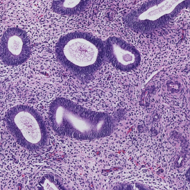
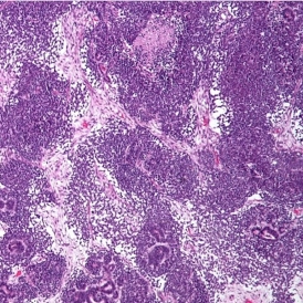
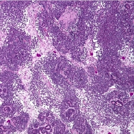
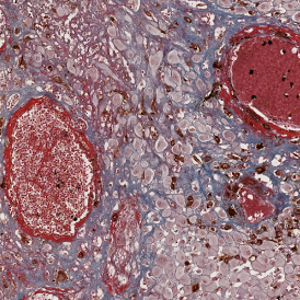
TissueGnostics provides advanced solutions for whole-slide imaging and image analysis in biological and clinical research. Our products help researchers to scan and analyze complex tissue samples, enabling more detailed insights into tissue structure, cellular interactions, and spatial cell landscape.


Keep in Touch

Be the first to hear about the latest scientific updates