


Analysis of cyclic stain and bleach samples
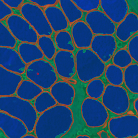
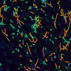
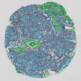
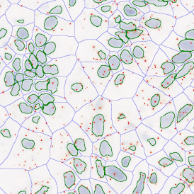
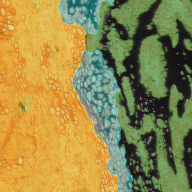
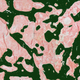
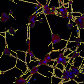
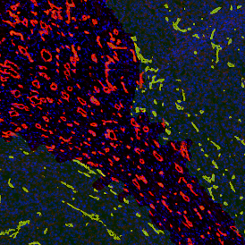
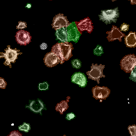
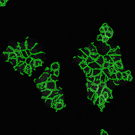
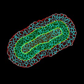
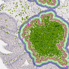
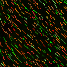
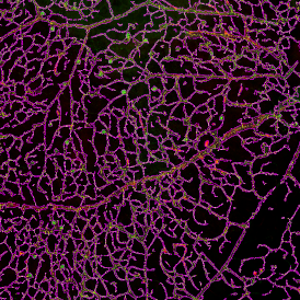
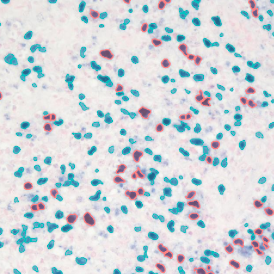
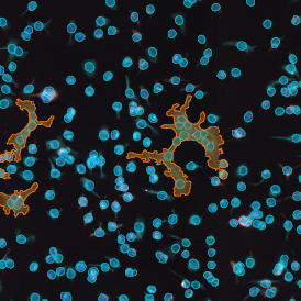
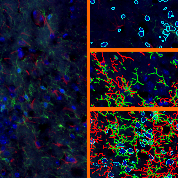
Multi-epitope ligand cartography (MELC) is a technology using samples subjected to cycles of fluorescent staining, imaging and photobleaching. Each cycle can use an antibody for a different protein. The result is a set of images of the distributions of many proteins for the same samples.
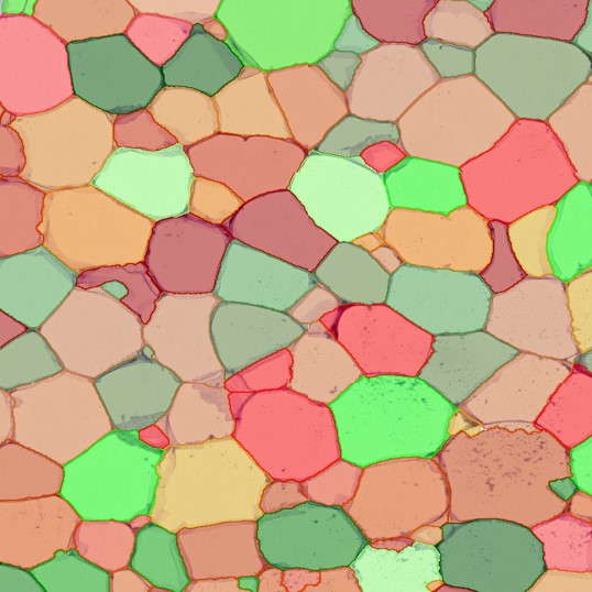
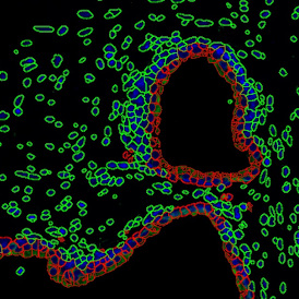
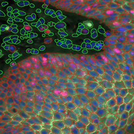
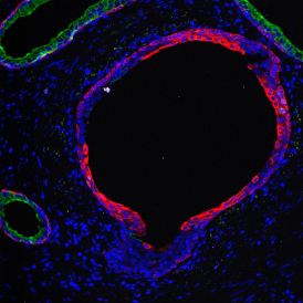
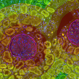
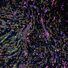
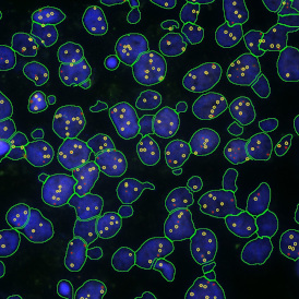
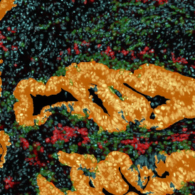
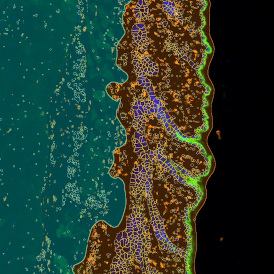
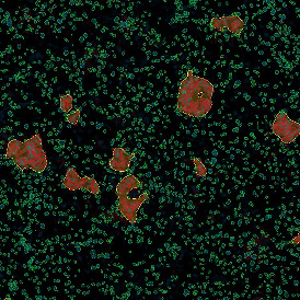
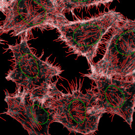
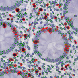
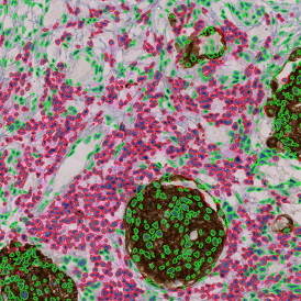
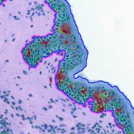
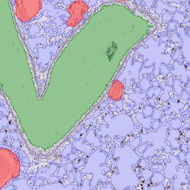
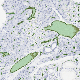
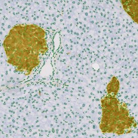
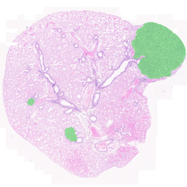
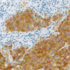
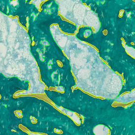
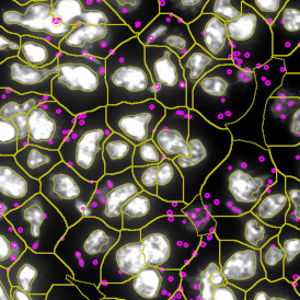
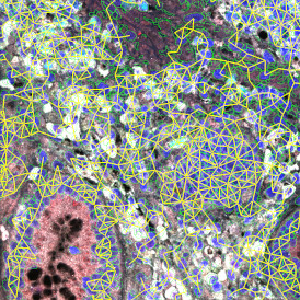
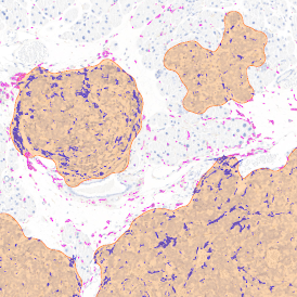
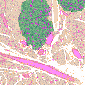
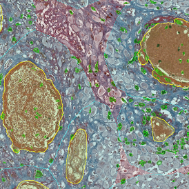
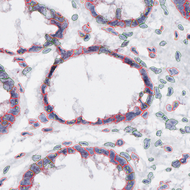
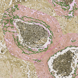
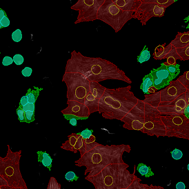
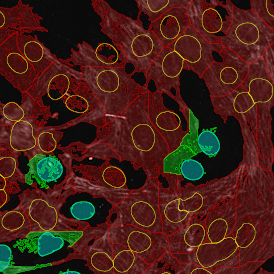
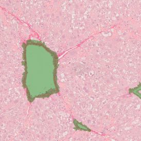
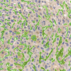
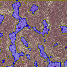
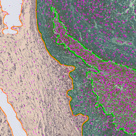
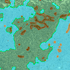
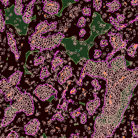
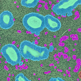
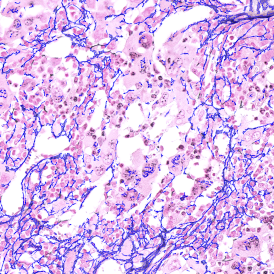
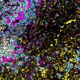
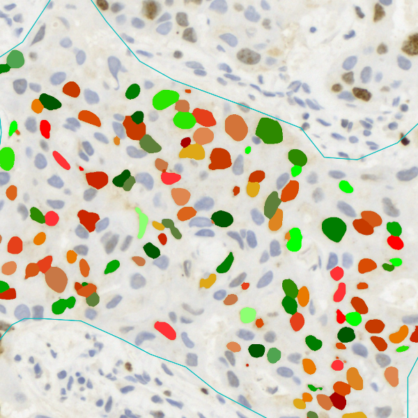
Tumor areas are detected using cytokeratin+ cells and their density. Blood vessels are detected using CD31+ cells. The distance of tumor cells from blood vessels within the tumor areas can then be measured. A specialized APP was established to analyse a MELC sample of skin from a melanoma patient. The aim was to quantify the expression of 90 proteins in close proximity to Melan-A positive areas. Some of the resulting images are shown below. StrataQuest is ideal to fully address the immense potential of such samples with automated context analysis.

The possibilities of the combination of cyclic stain-bleach methods and StrataQuest context-based analysis are almost endless. Which protein increases in its expression density and which one decreases in relation to its distance to:
- Normal melanocytes,
- Melanoma cells,
- Blood vessels,
- Inflammation sites,
- Sweat glands,
- Hair follicles,
- Basal membrane,
- Langerhans cells,
- Intraepithelial T-cells,
- Regulatory T-cells
- ...